组成图示
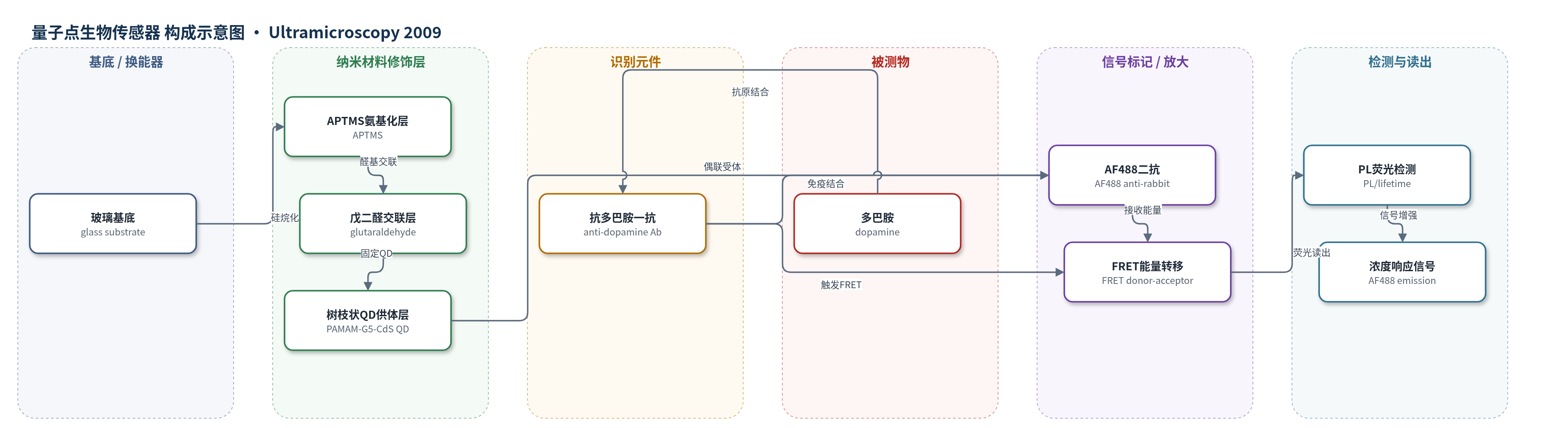
点击图片查看大图 · 依据论文自动绘制
传感器类型
量子点生物传感器
检测对象
多巴胺(dopamine, DA);样品基质为PBS/Tris缓冲液ELISA体系(未明确血清、脑脊液等临床基质)
检测原理
该检测基于免疫夹心与FRET光学换能。PAMAM-G5包封CdS QD作为供体,AF488标记的山羊抗兔IgG作为受体,兔抗多巴胺一抗识别多巴胺。多巴胺与一抗结合后,二抗通过免疫结合靠近一抗,使供体-受体距离和取向发生变化,FRET效率改变。多巴胺浓度升高时,抗原-抗体复合物增多,AF488受体发射增强,CdS QD供体发射同步降低,PL强度与荧光寿命随之变化。系统通过PL/OD读出AF488信号,实现多巴胺浓度响应。树枝状聚合物包封提高QD发光效率,免疫结合提供识别与信号放大。
检测灵敏度
测试浓度: 0–10 mM;0–5 mM内呈均匀变化
效应效果
在ELISA体系中,FRET效率达0.96,供体-受体距离约26 Å;AF488发射随多巴胺浓度增加而增强,供体发射同步降低,0–5 mM内变化均匀,10 mM仍增强。反应30 min后信号显著上升,随后增速下降,提示抗原-抗体结合主要发生在早期。玻璃表面经APTMS/戊二醛修饰后接触角由7.01°升至60.71°/53.81°,AFM显示粗糙度变化,证明表面固定可行。G5树枝状聚合物包封CdS QD发光效率比低代高约100倍。未报告选择性、稳定性、重现性、回收率及方法对比。作者认为其可发展为光学生物试剂盒/传感器,用于帕金森病多巴胺实时监测。
传感器的构成
- 基底:玻璃片(glass substrate),经Piranha溶液清洗,提供透明支撑与光学读出表面。
- 硅烷化层:3-氨基丙基三乙氧基硅烷(APTMS),在玻璃表面引入氨基,用于后续交联。
- 交联层:戊二醛(glutaraldehyde, GA),与APTMS氨基形成席夫碱,提供醛基固定树枝状聚合物。
- 量子点供体层:PAMAM树枝状聚合物(G5)包封CdS量子点(QD),作为FRET供体并固定于表面。
- 识别元件:兔抗多巴胺多克隆抗体(rabbit anti-dopamine polyclonal antibody),特异性识别多巴胺。
- 信号受体:AlexaFluor 488山羊抗兔IgG抗体(AF488 goat anti-rabbit IgG),作为FRET受体并发射荧光。
- 交联/还原剂:戊二醛(GA)与硼氢化钠(NaBH4),用于连接dendrimer-QD与抗体。
- 封闭/稳定剂:BSA、NGS、Triton X-100、焦亚硫酸钠(SMB),用于封闭非特异结合并稳定反应。
中文摘要
统一帕金森病评分量表(UPDRS)目前用于评估帕金森病,但主要依赖总体临床表现,难以定量判断病情进展,也无法实时指导药物剂量。多巴胺浓度是判断该病的关键因素,因此需要发展可定量检测多巴胺的生物试剂盒或生物传感器。本研究利用供体-受体间的Förster共振能量转移(FRET)现象,考察多巴胺浓度对能量转移的影响。以PAMAM树枝状聚合物(G5)包封的CdS量子点(QD)为供体,AlexaFluor 488标记的山羊抗兔IgG抗体为受体,通过ELISA/免疫荧光体系检测多巴胺。PL光谱和荧光寿命结果表明,随着多巴胺浓度增加,能量转移程度增强,AF488发射增强而供体发射降低。为构建光学传感器,玻璃表面经APTMS、戊二醛和dendrimer-QD修饰,并用接触角、ATR-FTIR和AFM表征表面润湿性、化学结构和形貌。研究为开发用于帕金森病多巴胺实时监测的光学生物传感器提供了基础。
英文摘要
The Unified Parkinson's Disease Rating Scale (UPDRS) is currently used to assess Parkinson's disease, and is a key method for determining the progression of disease based on the gross findings of patients. However, this method cannot quantify the extent of disease of patients, which means the administration of drugs cannot be determined on a real-time basis. Thalamotomy also causes discomfort and pain to the patients, and adversely affects treatment as it is performed following the onset of symptoms. Accordingly, the dopamine concentration, which is one of the key factors in determining this disease, needs to be detected quantitatively at ordinary times. Hence, the development of a bio-kit or a bio-sensor is essential for effectively prescribing the correct dopamine concentration in a customizable manner. In this study, the effect of dopamine level on this phenomenon was observed using the Forster resonance energy transfer (FRET) phenomenon generated between a donor and acceptor. By confirming the photoluminescence (PL) and lifetime data, it was demonstrated that the degree of energy transfer increased with increasing dopamine concentration. To apply this phenomenon to an optical sensor, a glass surface was modified with a quantum dot (QD)-encapsulated dendrimer, and analyzed using the contact angle and ATR-FTIR. The topology of surface was determined by an atomic force microscope (AFM).