组成图示
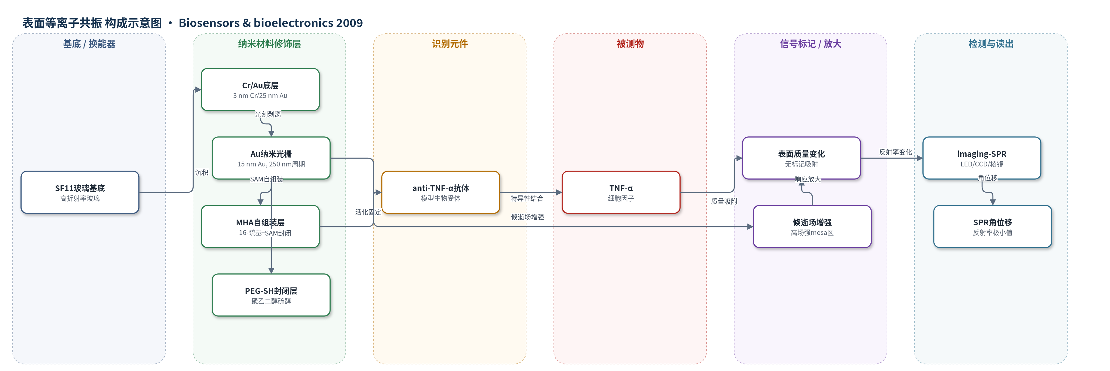
点击图片查看大图 · 依据论文自动绘制
传感器类型
表面等离子共振(SPR)生物传感器
检测对象
肿瘤坏死因子α(TNF-α);样品基质:PBS 缓冲液(磷酸盐缓冲液)
检测原理
该传感器采用 Kretschmann 构型 SPR 换能。TM 偏振光经棱镜激发金膜表面等离子体,共振角取决于金属-介质界面附近折射率。金纳米光栅扰动表面等离子体传播,使倏逝场在 mesa 及边缘集中,形成高场强区域。16-巯基十六酸 SAM 经 EDC/NHS 活化后固定 anti-TNF-α 抗体,PEG-SH 封闭非功能化区域。TNF-α 与抗体结合后,界面质量与折射率增加,导致 SPR 共振角位移。由于识别元件位于高场强区且三维可及性提高,相同结合量产生更大角位移,从而实现无标记、实时、信号放大的检测。
检测灵敏度
原文未报告 LOD、线性范围、灵敏度斜率及相关系数。
效应效果
实验以 anti-TNF-α/TNF-α 为模型体系,在 PBS 中进行成像 SPR 测量。纳米光栅 mesa 功能化表面给出最大 SPR 角位移;相比平面表面或均匀功能化纳米光栅,anti-TNF-α 吸附的角共振响应提高约 3 倍。严格耦合波模拟显示其角响应较平面几乎提高 3 倍,较均匀功能化纳米光栅大 40%。TNF-α 结合时,mesa 纳米光栅表面角位移平均增加约 8 倍,而 anti-TNF-α 吸附时约为 4–5 倍。PEG-SH 封闭降低非特异吸附,但边缘效应和工艺波动仍造成样本间变异。作者认为该图案化纳米光栅界面可用于高灵敏度集成 SPR 生物传感器和现场检测。
传感器的构成
- 基底/换能器:SF11 高折射率玻璃,作为 Kretschmann 棱镜耦合 SPR 的支撑与光路基底
- 粘附层:3 nm Cr,增强金层与玻璃结合
- 底层换能金属:25 nm Au,形成 SPR 金属-介质界面
- 纳米光栅层:15 nm Au,周期 250 nm、深 15 nm、占空比 50%,重分布倏逝场并增强高场区
- 识别固定层:16-巯基十六酸(16-mercaptohexadecanoic acid)自组装单分子层,提供羧基并经 EDC/NHS 活化后固定抗体
- 封闭层:聚乙二醇硫醇(PEG-SH)自组装单分子层,封闭未功能化区域,减少蛋白非特异吸附
- 识别元件:anti-TNF-α 抗体,特异性捕获 TNF-α
- 信号标记物:无外源标记,依靠表面质量/折射率变化产生 SPR 角位移
- 读出系统:imaging-SPR(SPRi-lab, Genoptic),LED 光源、棱镜与 CCD 相机测量反射率/角位移
中文摘要
本文报道了一种用于表面等离子共振(SPR)生物传感器的纳米光栅增强生物界面。作者提出将表面生物受体图案化固定于金纳米光栅的高电磁场区域,以提高 SPR 角响应灵敏度。该界面采用 Kretschmann 构型,通过电子束光刻、金属 lift-off 和自组装单分子层制备:在 SF11 玻璃/金基底上形成周期 250 nm、深 15 nm 的金纳米光栅,并用 16-巯基十六酸和聚乙二醇硫醇分别实现抗体固定与封闭。严格耦合波分析表明,将生物受体固定于高场强区域可增大 SPR 角位移。成像 SPR 实验以 anti-TNF-α 抗体为模型生物受体,结果显示功能化 mesa 的纳米光栅表面相比平面表面或均匀功能化纳米光栅表面,角共振响应提高约 3 倍;同时由于纳米光栅上生物受体可及性增加,TNF-α 的检测信号也增强。
英文摘要
In this report, nano-gratings with guided adsorption of biomolecules are investigated as new transducer elements or biointerfaces for surface plasmon resonance biosensor technologies. SPR biosensors are of particular interest due to the interaction between the electromagnetic fields and periodic nano-structures. In this article, sensitivity enhancement is demonstrated for a surface plasmon resonance interface, in a Kretschmann's configuration, featuring nano-gratings combined with nano-patterned immobilization of surface bioreceptors. The fabrication of this enhanced biointerface is demonstrated using a combination of metal lift-off and self-assembled monolayers. Rigorous coupled-wave analyses point to an increase in SPR angular response for the immobilization of surface bioreceptors onto areas of the nano-corrugated surface exhibiting high electromagnetic field intensity. Experimental measurements of the immobilization of anti-TNF-alpha antibody as a model bioreceptor using an imaging-SPR technique show a 3 times increase in angular resonance response from nano-grating surfaces with functionalized mesas compared to a planar surface or to a uniformly functionalized nano-grating surface. Furthermore, results also show an increased detection of TNF-alpha due to the increased accessibility to the adsorbed bioreceptors on the nano-gratings.