组成图示
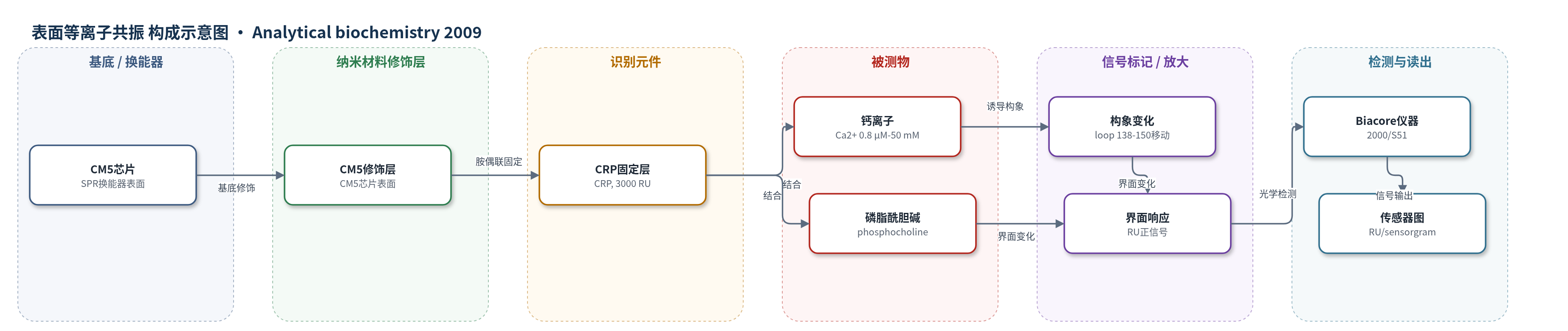
点击图片查看大图 · 依据论文自动绘制
传感器类型
表面等离子共振(SPR)生物传感器
检测对象
钙离子(Ca2+)、磷脂酰胆碱(phosphocholine);样品基质为缓冲液流动相(10 mM Hepes/100 mM NaCl/10 mM CaCl2/0.005% P-20,或无Ca2+及Mops/Mes/Na-acetate pH 5.0–8.0)。
检测原理
CRP经胺偶联固定于CM5芯片,形成SPR识别表面。注入Ca2+后,Ca2+与CRP两个独立位点结合;低亲和力位点结合诱导138–150环移入蛋白主体,引起显著构象变化,使界面折射率/质量分布改变,产生约为质量估算10倍的正向RU信号。稳态RU随Ca2+浓度呈双S形,用双位点模型拟合KD。phosphocholine结合依赖Ca2+,呈1:1可逆结合,无协同性,信号主要来自界面质量/折射率变化。SPR实时记录RU,扣除参考与空白后以稳态值全局非线性回归,无HCR/RCA等化学放大。
检测灵敏度
未报告检出限(LOD)、线性范围、灵敏度斜率或相关系数。
效应效果
CRP固定化表面稳定,约3000 RU,Ca2+解离快,无需再生。选择性上,仅二价离子有响应,约为Ca2+的一半,单价阳离子无信号;phosphocholine结合在1 mM EDTA下消失,呈钙依赖。定量误差:phosphocholine KD=5.0±3.3 μM;Ca2+高亲和力KD=0.03±0.011 mM,低亲和力KD=5.45±0.52 mM;高亲和力位点pKa=4.25±0.12,低亲和力位点拐点pH=5.93±0.12。未报告RSD、回收率及与ELISA/HPLC/qPCR对比。作者认为SPR可检测伴随构象变化的小分子/离子结合,并提示Ca2+生理调控CRP。
传感器的构成
- 基底/换能器:Biacore CM5 传感器芯片(SPR 换能器),提供表面等离子共振检测表面。
- 识别元件:C-reactive protein(CRP),经 amine coupling 固定于 CM5 芯片,约 3000 RU,作为结合 Ca2+ 与 phosphocholine 的受体。
- 反应介质:10 mM Hepes(pH 7.4)、100 mM NaCl、10 mM CaCl2、0.005% surfactant P-20;Ca2+ 实验使用无 Ca2+ 缓冲液或 50 mM Mops、50 mM Mes、50 mM Na-acetate(pH 5.0–8.0),用于维持蛋白活性并控制 Ca2+ 与 pH。
- 分析物:Ca2+(CaCl2 浓度系列)或 phosphocholine(0.16–100 μM),注入流动相与固定化 CRP 结合。
- 信号读出:Biacore 2000/S51 SPR 仪器,以 resonance units(RU)记录 sensorgrams,并通过稳态拟合获得 KD。
中文摘要
本研究利用表面等离子共振(SPR)生物传感器表征钙离子(Ca2+)与C反应蛋白(CRP)的相互作用。将CRP固定于传感器芯片上,依次注入不同浓度的Ca2+或磷脂酰胆碱(phosphocholine)。Ca2+结合产生的信号比仅按其分子量估算高约10倍,作者将其归因于Ca2+结合诱导的构象变化。稳态数据可用两个独立结合位点模型拟合:高亲和力位点KD为0.03 mM,低亲和力位点KD为5.45 mM。两个位点的pH依赖性不同,从而可将其对应到CRP三维结构中的不同Ca2+配位位点。磷脂酰胆碱与CRP的结合呈钙依赖性,在10 mM Ca2+下KD为5 μM,且未观察到协同性。结果表明,SPR生物传感器能够在小分子或离子结合伴随显著构象变化时,有效检测并定量其与固定化蛋白的相互作用,为理解CRP功能及Ca2+在生理条件下的调控作用提供了依据。
英文摘要
The interactions between Ca2+ and C-reactive protein (CRP) have been characterized using a surface plasmon resonance (SPR) biosensor. The protein was immobilized on a sensor chip, and increasing concentrations of Ca2+ or phosphocholine were injected. Binding of Ca2+ induced a 10-fold higher signal than expected from the molecular weight of Ca2+. It was interpreted to result from the conformational change that occurs on binding of Ca2+. Two sites with different characteristics were distinguished: a high-affinity site with K(D)=0.03 mM and a low-affinity site with K(D)=5.45 mM. The pH dependencies of the two Ca2+ interactions were different and enabled the assignment of the different sites in the three-dimensional structure of CRP. There was no evidence for cooperativity in the phosphocholine interaction, which had K(D)=5 microM at 10 mM Ca2+. SPR biosensors can clearly detect and quantify the binding of very small molecules or ions to immobilized proteins despite the theoretically very low signals expected on binding, provided that significant conformational changes are involved. Both the interactions and the conformational changes can be characterized. The data have important implications for the understanding of the function of CRP and suggest that Ca2+ is an efficient regulator under physiological conditions.