组成图示
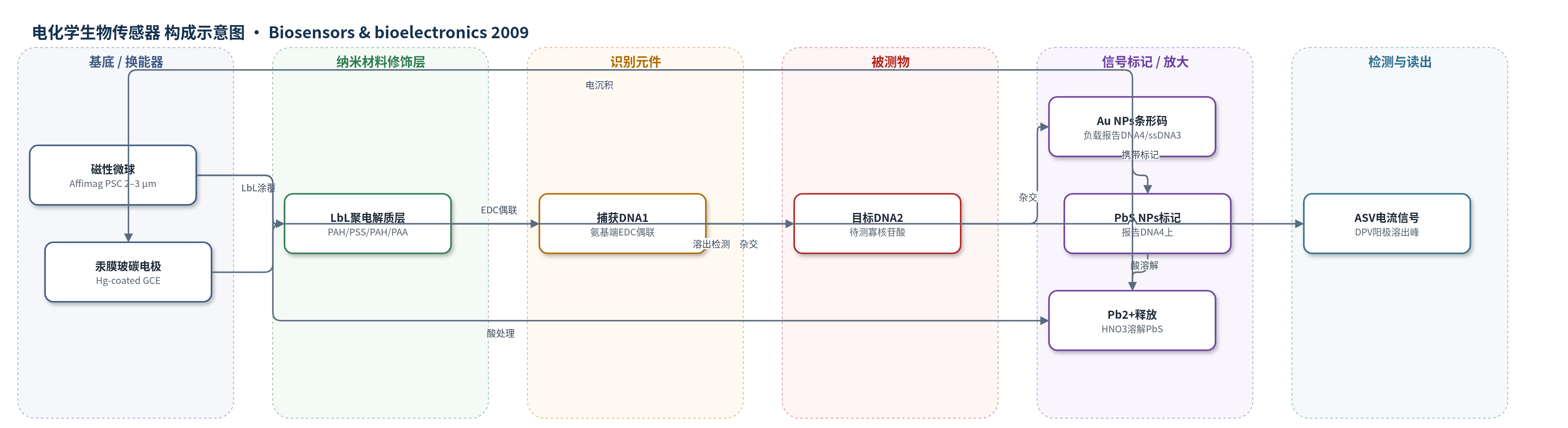
点击图片查看大图 · 依据论文自动绘制
传感器类型
电化学生物传感器
检测对象
目标 DNA(target DNA,寡核苷酸);样品基质为 PBS 缓冲液/溶液(未涉及血清等实际基质)
检测原理
磁性微球经 PAH/PSS/PAH/PAA 层层自组装后表面羧基增多,通过 EDC 偶联固定更多氨基捕获 DNA1。目标 DNA2 与捕获 DNA1 杂交后,其另一端与负载 Au NPs 生物条形码的信号探针杂交;每个 Au NP 可携带多条报告 DNA4 和 ssDNA3,PbS NPs 标记在报告 DNA4 上,使每个目标 DNA 对应多个 PbS 标记。杂交完成后用 1.0 M HNO3 溶解 PbS,释放的 Pb2+ 浓度与目标 DNA 浓度成正比。Pb2+ 在汞膜玻碳电极上阴极预富集,随后差分脉冲阳极溶出伏安法(DPV/ASV)产生阳极溶出峰,峰电流随目标 DNA 浓度增大。信号放大来自 LbL 增加捕获 DNA、Au NPs 生物条形码增加 PbS 标记以及 ASV 预富集。
检测灵敏度
LOD: 5.0 × 10⁻¹⁵ M;线性范围: 2.0 × 10⁻¹⁴ M–1.0 × 10⁻¹² M(Y = 1.5215X −0.0070X2,X 为 10−14 M,Y 为 10−6 A,n = 12,R^2 = 0.9952);低浓度线性范围: 2.0 × 10⁻¹⁴ M–1.0 × 10⁻¹³ M(Y = 1.7128X + 0.8829,X 为 10−14 M,Y 为 10−6 A,n = 6,r = 0.9925)
效应效果
该传感器对完全互补目标 DNA 产生明显铅阳极溶出峰,对双碱基错配 DNA 的响应显著降低,对非互补 DNA 几乎无响应,表明杂交识别具有良好选择性。铅离子测定相对标准偏差(RSD)为 5.6%(n=7),重现性良好。信号 DNA 探针在冰箱中至少稳定 1 个月,磁性微球载体在 4 ℃ 缓冲液中至少稳定 8 h,电化学信号保持稳定。作者认为该三明治杂交体系兼具高灵敏度、良好选择性和稳定性,可作为其他 DNA 生物检测方法的替代方案,其灵敏度与基于金属纳米颗粒的 DNA 杂交检测相当。
传感器的构成
- 磁性载体:Affimag PSC 磁性微球(2.0–3.0 μm),作为固相捕获基底并便于磁分离
- 聚电解质修饰层:PAH/PSS/PAH/PAA 四层(LbL),增加表面羧基以固定更多捕获 DNA
- 识别元件:氨基捕获 DNA1(5'-NH2-GCG-CGA-ACC-GTA-TA-3'),经 EDC 偶联到 PAA 羧基,捕获目标 DNA
- 信号纳米标记:Au NPs(约 20 nm)生物条形码,负载报告 DNA4 与 ssDNA3,杂交到目标 DNA 另一端并放大信号
- 电化学标记物:PbS NPs(约 10 nm),标记在报告 DNA4 上,作为可溶解释放 Pb2+ 的信号源
- 换能器电极:汞膜修饰玻碳电极(Hg-coated GCE),用于阳极溶出伏安法(ASV)检测 Pb2+;辅助电极为 Pt 丝,参比电极为 Ag/AgCl
- 读出介质:1.0 M HNO3 溶解 PbS 释放 Pb2+,0.1 M HAc–NaAc(pH 5.3)支持电解质,ASV 读取阳极溶出峰
中文摘要
本文构建了一种基于磁性微球、金纳米颗粒(Au NPs)生物条形码和硫化铅纳米颗粒(PbS NPs)放大的三明治电化学生物传感器。磁性微球经四层聚电解质层层自组装(LbL)后增加表面羧基,从而固定更多氨基捕获 DNA。捕获 DNA 与目标 DNA 一端杂交,目标 DNA 另一端与标记 Au NPs 的信号 DNA 探针杂交;Au NPs 负载生物条形码,PbS NPs 作为目标寡核苷酸标记物。杂交后用硝酸溶解 PbS 释放铅离子,并以阳极溶出伏安法(ASV)检测。LbL 修饰提高捕获 DNA 负载量,生物条形码增加 Au NPs 数量,ASV 进一步放大信号。最佳条件下,目标 DNA 在 2.0×10⁻¹⁴ M 至 1.0×10⁻¹² M 线性,检出限 5.0×10⁻¹⁵ M,传感器选择性和灵敏度良好。
英文摘要
A novel and sensitive sandwich electrochemical biosensor based on the amplification of magnetic microbeads and Au nanoparticles (NPs) modified with bio bar code and PbS nanoparticles was constructed in the present work. In this method, the magnetic microspheres were coated with 4 layers polyelectrolytes in order to increase carboxyl groups on the surface of the magnetic microbeads, which enhanced the amount of the capture DNA. The amino-functionalized capture DNA on the surface of magnetic microbeads hybridized with one end of target DNA, the other end of which was hybridized with signal DNA probe labelled with Au NPs on the terminus. The Au NPs were modified with bio bar code and the PbS NPs were used as a marker for identifying the target oligoncleotide. The modification of magnetic microbeads could immobilize more amino-group terminal capture DNA, and the bio bar code could increase the amount of Au NPs that combined with the target DNA. The detection of lead ions performed by anodic stripping voltammetry (ASV) technology further improved the sensitivity of the biosensor. As a result, the present DNA biosensor showed good selectivity and sensitivity by the combined amplification. Under the optimum conditions, the linear relationship with the concentration of the target DNA was ranging from 2.0 x 10(-14) M to 1.0 x 10(-12)M and a detection limit as low as 5.0 x 10(-15)M was obtained.