组成图示
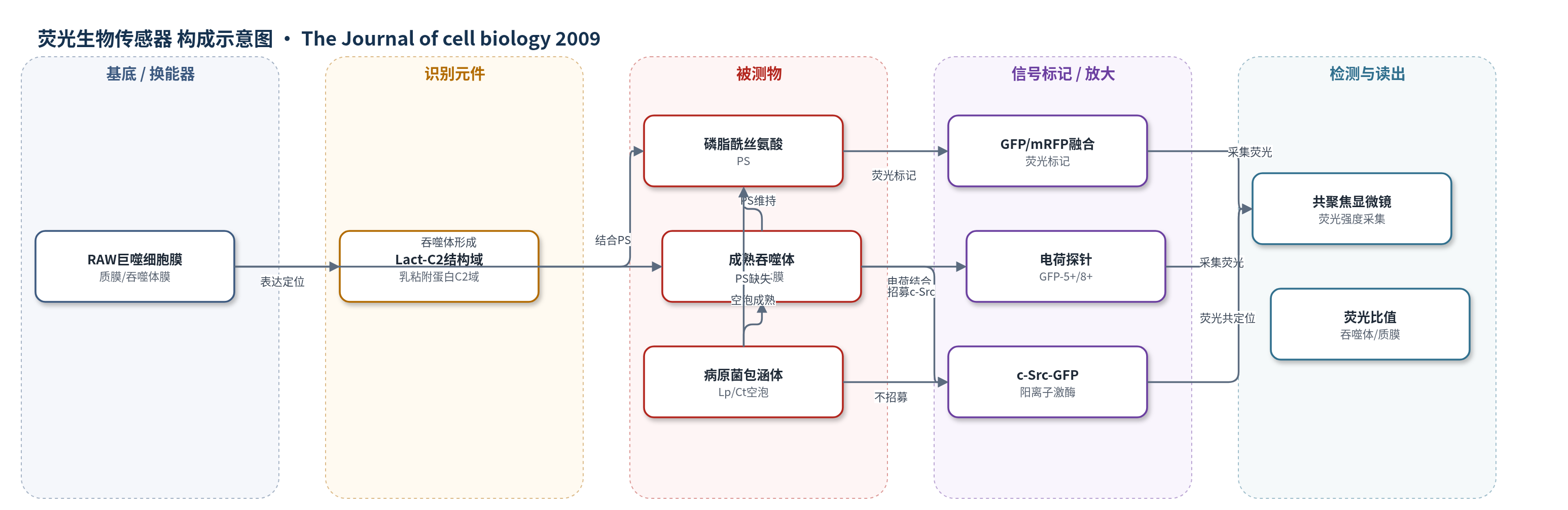
点击图片查看大图 · 依据论文自动绘制
传感器类型
荧光生物传感器
检测对象
磷脂酰丝氨酸(phosphatidylserine, PS);样品基质:RAW264.7 巨噬细胞活细胞质膜、吞噬体膜及 Legionella pneumophila/Chlamydia trachomatis 感染形成的包涵体膜
检测原理
该检测基于遗传编码荧光生物传感器:乳粘附蛋白 C2 结构域(Lact-C2)与 GFP 或 mRFP 融合表达于 RAW264.7 巨噬细胞中。Lact-C2 对磷脂酰丝氨酸(PS)具有立体选择性,可结合暴露于膜胞质侧的 PS。当吞噬体膜上 PS 密度升高时,更多探针结合并产生更强荧光;PS 被清除时荧光降低。通过旋转盘共聚焦显微镜采集吞噬体膜与未参与质膜的荧光强度,计算比值,从而动态反映 PS 分布。多阳离子脂锚定探针(GFP-8+、GFP-5+)则依据膜表面负电荷结合,用于评估 PS 维持的表面电位及其对 c-Src 等阳离子蛋白的招募。方法无外源酶或核酸放大,信号直接来自蛋白-脂质结合与荧光标记。
检测灵敏度
原文未报告 LOD、线性范围、灵敏度斜率或相关系数。
效应效果
与 annexin-V 和抗 PS 抗体相比,Lact-C2 探针无需固定、通透或高钙条件,且对 PS 更具立体选择性;文中显示抗 PS 抗体可结合 PA 和 PI,annexin-V 可结合 PA 与 PG,说明传统试剂特异性不足。FM4-64 标记内体膜与 Lact-C2 的 Manders 共定位系数为 0.933,支持内体融合向吞噬体递送 PS。GFP-Lact-C2 在吞噬体封闭后至少 1 h 持续存在,5 min 至 1 h 密度基本恒定,而质膜标记 mRFP-Palm 和 GT46 明显减少。病原菌 Legionella pneumophila 与 Chlamydia trachomatis 的成熟空泡缺乏 PS,5+ 探针和 c-Src 均不能有效招募。作者认为 PS 是吞噬体成熟和杀菌的关键膜成分。
传感器的构成
- 细胞膜基底:RAW264.7 巨噬细胞质膜与吞噬体膜,提供 PS 暴露的胞质侧界面
- 识别元件:乳粘附蛋白 C2 结构域(Lact-C2),选择性结合磷脂酰丝氨酸(PS)
- 荧光标记物:绿色荧光蛋白(GFP)或单体红色荧光蛋白(mRFP)融合于 Lact-C2,作为 PS 信号标记
- 辅助电荷探针:GFP-8+、GFP-5+、GFP-2+ 等多阳离子脂锚定探针,用于评估膜表面电荷
- 膜标记对照:mRFP-Palm 与 GT46,用于监测质膜成分在吞噬体中的保留与清除
- 信号读出:旋转盘共聚焦显微镜采集 GFP/mRFP 荧光强度并计算吞噬体/质膜比值
中文摘要
在吞噬作用中,活化膜的磷脂酰肌醇含量及其相关表面电荷急剧下降,从而吸引多阳离子蛋白。质膜胞质侧富含磷脂酰丝氨酸(PS),但缺乏合适探针使该磷脂在吞噬过程中的命运难以研究。本研究使用近期开发的荧光生物传感器监测吞噬体形成与成熟过程中 PS 的分布和动态。与多磷酸肌醇不同,PS 在吞噬体封闭后仍持续存在于吞噬体膜上,即使其他质膜成分已被清除。通过与内体和溶酶体融合,吞噬体维持较高 PS 水平,并足以招募 c-Src 等阳离子蛋白。含有军团菌和沙眼衣原体的吞噬空泡因偏离内体-溶酶体成熟途径而缺乏 PS,表面电荷很低,且不能招募 c-Src。这些结果强调了 PS 在吞噬体成熟和微生物杀灭中的功能。
英文摘要
During phagocytosis, the phosphoinositide content of the activated membrane decreases sharply, as does the associated surface charge, which attracts polycationic proteins. The cytosolic leaflet of the plasma membrane is enriched in phosphatidylserine (PS); however, a lack of suitable probes has precluded investigation of the fate of this phospholipid during phagocytosis. We used a recently developed fluorescent biosensor to monitor the distribution and dynamics of PS during phagosome formation and maturation. Unlike the polyphosphoinositides, PS persists on phagosomes after sealing even when other plasmalemmal components have been depleted. High PS levels are maintained through fusion with endosomes and lysosomes and suffice to attract cationic proteins like c-Src to maturing phagosomes. Phagocytic vacuoles containing the pathogens Legionella pneumophila and Chlamydia trachomatis, which divert maturation away from the endolysosomal pathway, are devoid of PS, have little surface charge, and fail to recruit c-Src. These findings highlight a function for PS in phagosome maturation and microbial killing.