组成图示
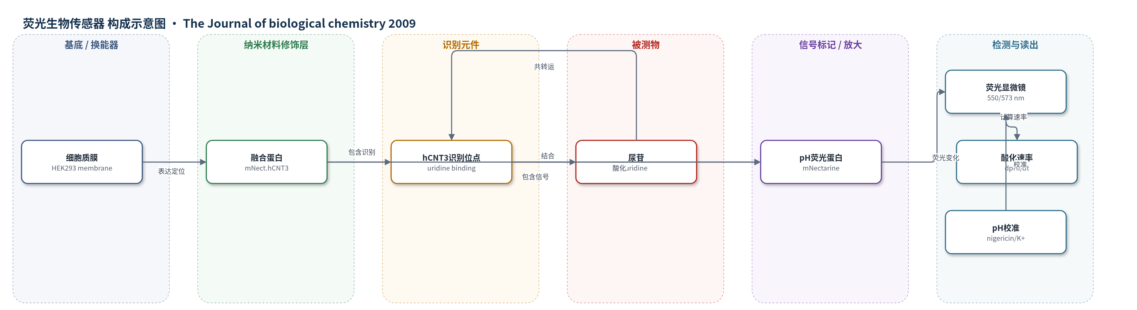
点击图片查看大图 · 依据论文自动绘制
传感器类型
荧光生物传感器
检测对象
尿苷(uridine)及其伴随 H+(proton)共转运;样品基质:HEK293 细胞培养液/MBSS 缓冲液(pH 5.5,含或不含 Na+)
检测原理
mNectarine(mNect)融合于 hCNT3 胞质 N 端,使 pH 敏感荧光基团位于转运蛋白胞内表面。酸性 MBSS 中,hCNT3 结合尿苷并介导 H+ 与尿苷共转运,导致 mNect 附近胞内 pH 下降。mNect 荧光强度随 pH 变化(pKa' 6.9),550 nm 激发下酸化使 573 nm 发射降低。先用 nigericin/高钾缓冲液钳制并校准 pHi,再加入尿苷,比较前后 20 s 的 dpHi/dt 差值,乘以 HEK293 胞质缓冲容量得到 H+ 通量。尿苷浓度升高时酸化速率增大,呈 Michaelis-Menten 动力学,从而实时反映 H+/尿苷共转运活性。
检测灵敏度
未报告 LOD、线性范围、灵敏度斜率或 R^2;报告 Km: 72 ± 24 μM(n = 3)
效应效果
pH 6.5–7.5 钳制下,mNect.hCNT3 报告胞质 pH 偏差 0.038 ± 0.005(n=24),BCECF 为 0.013 ± 0.003(n=12)。加入 0.5 mM 尿苷后,mNect.hCNT3 的 H+ 通量 1.08 ± 0.10 mM/min,阴性对照 -0.02 ± 0.1 mM/min(p<0.0001)。Km 72 ± 24 μM,接近卵母细胞 110 ± 10 与 62.4 ± 5.4 μM。含/无 Na+ 通量 1.18 ± 0.32 与 1.09 ± 0.11 mM/min(p=0.81)。方法无需放射性底物,可实时自参照检测核苷转运。
传感器的构成
- 基底/定位层:HEK293 细胞质膜(plasma membrane),承载融合蛋白并分隔胞外与胞内 pH。
- 转运蛋白锚定层:hCNT3(human concentrative nucleoside transporter 3),跨膜转运蛋白,将 mNect 锚定于胞内表面。
- 识别元件:hCNT3 核苷结合位点,识别并结合尿苷(uridine),介导 H+/尿苷共转运。
- 信号换能元件:mNectarine(mNect),pH 敏感单体红色荧光蛋白(mRFP),pKa' 6.9,荧光强度随胞内 pH 变化。
- 阴性对照元件:mNect.hCNT3-F563C,hCNT3 失活突变体融合 mNect,用于排除非转运相关酸化。
- 校准/参考体系:nigericin/高钾 pH 钳制缓冲液,用于将 mNect 荧光转换为 pHi;BCECF 作为传统 pH 染料对照。
- 读出系统:倒置荧光显微镜/显微光度计,550 nm 激发、573 nm 发射,记录荧光并计算 dpHi/dt。
中文摘要
人浓缩型核苷转运体 hCNT3 可介导 Na+/核苷和 H+/核苷共转运。本文报道一种在培养哺乳动物细胞中监测 H+/尿苷共转运的新方法,并开发、表征了 pH 敏感单体红色荧光蛋白变体 mNectarine。将 mNectarine 融合至 hCNT3 N 端形成 mNect.hCNT3,可在 hCNT3 胞内表面测量 pH。在表达 mNect.hCNT3 或失活突变体 mNect.hCNT3-F563C 的 HEK293 细胞中监测荧光;游离胞质 mNect、mNect.hCNT3 与传统 pH 染料 BCECF 在 pH 钳制细胞中报告胞质 pH 相似。在允许 H+ 偶联转运的 pH 5.5、无 Na+ 和含 Na+ 条件下,仅 mNect.hCNT3 表达细胞在加入 0.5 mM 尿苷后酸化速率增加,首次直接证明 H+ 偶联尿苷转运。pH 5.5 时,有无 Na+ 的尿苷转运速率(偶联 H+ 通量)无显著差异(1.09 ± 0.11 或 1.18 ± 0.32 mM/min),提示酸性含 Na+ 条件下每分子尿苷转运 1 个 Na+ 和 1 个 H+,酸性无 Na+ 条件下仅转运 1 个 H+。该融合蛋白为核苷转运提供了简单、自参照且有效的监测手段,可推广至其他 H+ 偶联转运蛋白活性检测。
英文摘要
Human concentrative nucleoside transporter, hCNT3, mediates Na+/nucleoside and H+/nucleoside co-transport. We describe a new approach to monitor H+/uridine co-transport in cultured mammalian cells, using a pH-sensitive monomeric red fluorescent protein variant, mNectarine, whose development and characterization are also reported here. A chimeric protein, mNectarine fused to the N terminus of hCNT3 (mNect.hCNT3), enabled measurement of pH at the intracellular surface of hCNT3. mNectarine fluorescence was monitored in HEK293 cells expressing mNect.hCNT3 or mNect.hCNT3-F563C, an inactive hCNT3 mutant. Free cytosolic mNect, mNect.hCNT3, and the traditional pH-sensitive dye, BCECF, reported cytosolic pH similarly in pH-clamped HEK293 cells. Cells were incubated at the permissive pH for H(+)-coupled nucleoside transport, pH 5.5, under both Na(+)-free and Na(+)-containing conditions. In mNect.hCNT3-expressing cells (but not under negative control conditions) the rate of acidification increased in media containing 0.5 mm uridine, providing the first direct evidence for H(+)-coupled uridine transport. At pH 5.5, there was no significant difference in uridine transport rates (coupled H+ flux) in the presence or absence of Na+ (1.09 +/- 0.11 or 1.18 +/- 0.32 mm min(-1), respectively). This suggests that in acidic Na(+)-containing conditions, 1 Na+ and 1 H+ are transported per uridine molecule, while in acidic Na(+)-free conditions, 1 H+ alone is transported/uridine. In acid environments, including renal proximal tubule, H+/nucleoside co-transport may drive nucleoside accumulation by hCNT3. Fusion of mNect to hCNT3 provided a simple, self-referencing, and effective way to monitor nucleoside transport, suggesting an approach that may have applications in assays of transport activity of other H(+)-coupled transport proteins.