组成图示
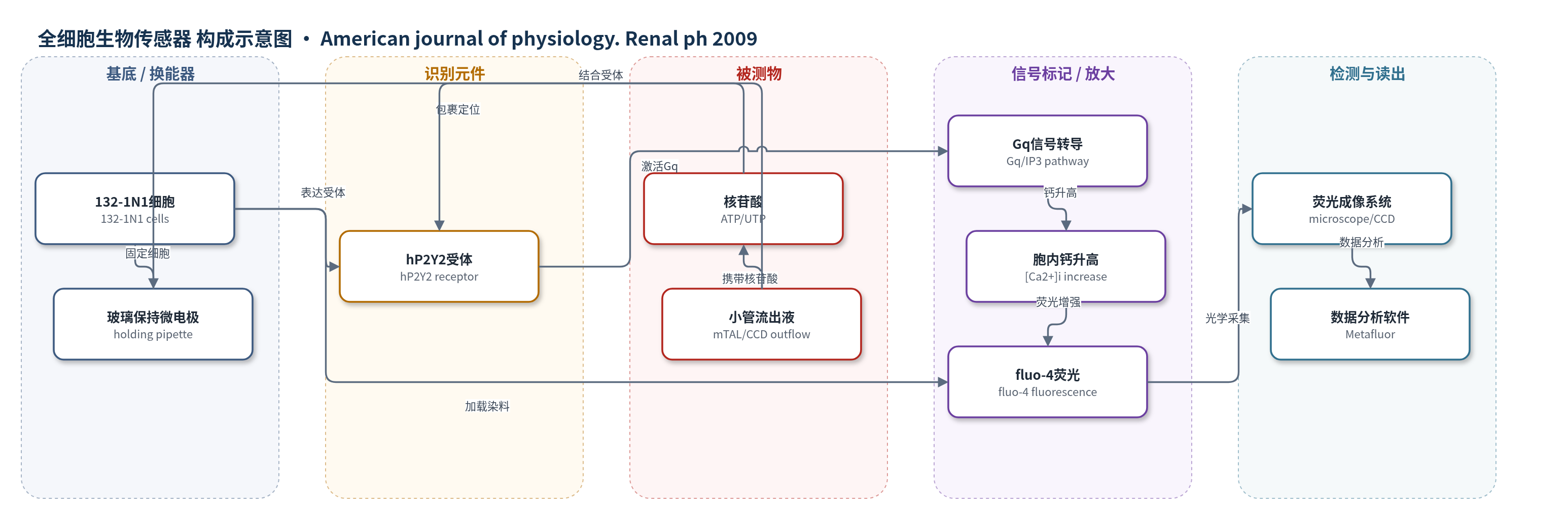
点击图片查看大图 · 依据论文自动绘制
传感器类型
全细胞生物传感器
检测对象
核苷酸(nucleotides,ATP/UTP),样品基质为小鼠髓质粗升支(mTAL)与皮质集合管(CCD)的管腔流出液/小管灌注液(tubular outflow/perfusate)
检测原理
该传感器以稳定表达人源 P2Y2 受体(hP2Y2)的 132-1N1 细胞为全细胞生物传感器。当肾小管流出液中的 ATP 或 UTP 与细胞膜 hP2Y2 受体结合时,受体激活 Gq 蛋白,经磷脂酶 C/IP3 途径促使内质网释放 Ca2+,导致胞内 Ca2+ 浓度升高。fluo-4 钙指示剂与 Ca2+ 结合后荧光增强,倒置显微镜在 488 nm 激发、>510 nm 发射下记录相对荧光变化。浓度响应显示,≥0.2 μM UTP/ATP 即可引起显著 [Ca2+]i 升高,EC50 分别为 UTP 0.26 μM、ATP 0.4 μM;1 μM UTP 的 1 s 短脉冲也能被检测。未转染 P2 受体的 132-1N1 细胞无响应,流速本身不引起信号,因此荧光变化主要反映管腔核苷酸浓度变化。
检测灵敏度
EC50 UTP: 0.26 μM;EC50 ATP: 0.4 μM;显著响应阈值: ≥0.2 μM UTP/ATP;短脉冲检测: 1 μM UTP, 1 s
效应效果
该传感器具有受体依赖性:未转染 132-1N1 细胞对 ATP/UTP 无 [Ca2+]i 响应,hP2Y2-132-1N1 细胞对 ≥0.2 μM UTP/ATP 显著响应,1 μM UTP 1 s 短脉冲可重复检测;流速本身和 10 nM AVP 均不引起生物传感器信号。静息 mTAL 8/8 实验在流出液中记录到自发核苷酸信号,移开即消失。AVP 在 mTAL 的 11 个合格实验中 7 个出现信号,共 20 次爆发;dDAVP 11 个中 8 个出现,共 16 次;AVP/dDAVP 首次信号延迟 99±39 s。CCD 7/7 出现信号,共 11 次,延迟 105±47 s。估算峰值管腔核苷酸浓度约 0.2–0.3 μM,与微穿刺水平相近。作者认为其可原位监测肾小管核苷酸分泌,揭示 AVP 的局部嘌呤能反馈。
传感器的构成
- 细胞基底/换能平台:132-1N1 细胞(132-1N1 cells)作为全细胞生物传感器平台,稳定表达 hP2Y2 受体并介导 Ca2+ 信号转导。
- 识别元件:人源 P2Y2 受体(hP2Y2)表达于 132-1N1 细胞膜,结合 ATP/UTP 并激活 Gq 通路。
- 信号标记物:fluo-4 钙荧光指示剂(fluo-4-AM 加载)随胞内 Ca2+ 升高产生荧光增强。
- 样品接触/定位组件:火抛光玻璃保持微电极(holding pipette)吸附 2–4 个 hP2Y2-132-1N1 细胞簇,置于肾小管流出液中。
- 光学读出系统:倒置显微镜(Axiovert 100TV)、单色器(Polychrome IV)、CCD 相机(MicroMax)与 Metamorph/Metafluor 软件,488 nm 激发、>510 nm 发射。
- 样品流路/灌注组件:同心玻璃微管灌注系统(perfusion pipettes)输送肾小管灌注液,使小管流出液包裹生物传感器细胞;fura-2 钾盐作为流动示踪剂用于定位。
- 培养/实验介质:DMEM + 10% FBS + 2 mM 谷氨酰胺(无核黄素)+ 青霉素/链霉素用于细胞培养;HEPES 缓冲生理溶液用于 37°C 实验。
中文摘要
细胞外核苷酸是局部、短寿命的信号分子,可通过管腔侧和基底外侧 P2 受体抑制肾小管转运。肾上皮本身能够释放核苷酸,但其释放机制及受刺激条件仍不清楚。本研究在完整、灌注的小鼠髓质粗升支(mTAL)和皮质集合管(CCD)中研究核苷酸分泌现象。作者将可记录小管流出液中核苷酸的生物传感器置于小管出口,同时用 fluo-4 同步测量生物传感器细胞与肾小管的胞内 Ca2+ 浓度。结果显示,静息灌注 mTAL 存在自发小管核苷酸分泌。在该制备中,10 nM 血管加压素(AVP)和 1-脱氨-8-D-精氨酸血管加压素(dDAVP)诱导强烈的 [Ca2+]i 振荡;AVP 在 CCD 中则诱导较大、缓慢且短暂的 [Ca2+]i 升高。重要的是,AVP/dDAVP 可触发 mTAL 小管核苷酸分泌。加入 AVP/dDAVP 后,生物传感器在小管灌注液中记录到核苷酸爆发,对应小管核苷酸浓度约 0.2–0.3 μM。CCD 经 AVP 刺激后也观察到非常相似的反应。因此,AVP 以爆发样模式刺激小管核苷酸分泌,峰值小管核苷酸浓度处于低微摩尔范围。作者推测,局部核苷酸信号是肾小管转运激素调控的内在反馈元件。
英文摘要
Extracellular nucleotides are local, short-lived signaling molecules that inhibit renal tubular transport via both luminal and basolateral P2 receptors. Apparently, the renal epithelium itself is able to release nucleotides. The mechanism and circumstances under which nucleotide release is stimulated remain elusive. Here, we investigate the phenomenon of nucleotide secretion in intact, perfused mouse medullary thick ascending limb (mTAL) and cortical collecting duct (CCD). The nucleotide secretion was monitored by a biosensor adapted to register nucleotides in the tubular outflow. Intracellular Ca(2+) concentration ([Ca(2+)](i)) was measured simultaneously in the biosensor cells and the renal tubule with fluo 4. We were able to identify spontaneous tubular nucleotide secretion in resting perfused mTAL. In this preparation, 10 nM AVP and 1-desamino-8-D-arginine vasopressin (dDAVP) induced robust [Ca(2+)](i) oscillations, whereas AVP in the CCD induced large, slow, and transient [Ca(2+)](i) elevations. Importantly, we identify that AVP/dDAVP triggers tubular secretion of nucleotides in the mTAL. After addition of AVP/dDAVP, the biosensor registered bursts of nucleotides in the tubular perfusate, corresponding to a tubular nucleotide concentration of approximately 0.2-0.3 microM. A very similar response was observed after AVP stimulation of CCDs. Thus AVP stimulated tubular secretion of nucleotides in a burst-like pattern with peak tubular nucleotide concentrations in the low-micromolar range. We speculate that local nucleotide signaling is an intrinsic feedback element of hormonal control of renal tubular transport.