组成图示
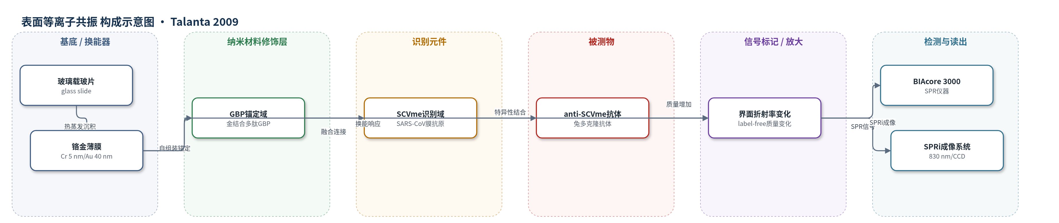
点击图片查看大图 · 依据论文自动绘制
传感器类型
表面等离子共振(SPR)生物传感器
检测对象
anti-SCVme 抗体(anti-SCVme antibody,兔抗 SARS-CoV 膜包膜抗原多克隆抗体);样品基质:PBS(磷酸盐缓冲液,pH 7.4),用于 SARS 诊断。
检测原理
GBP-E-SCVme 融合蛋白通过金结合多肽(GBP)域与金表面直接自组装,无需化学修饰,使 SCVme 抗原域以适当取向暴露于流动液。当样品中的 anti-SCVme 抗体流过芯片时,抗体与 SCVme 域发生特异性抗原—抗体结合,使金表面附近质量增加并改变局部折射率。该界面变化调制表面等离子共振条件,导致共振角或反射光强度变化。BIAcore 3000 以共振单位(RU)实时读出信号,SPRi 则以 830 nm 斑点强度成像。anti-SCVme 浓度越高,结合量越大,SPR 响应越大;BSA 封闭可抑制非特异结合,提高选择性。该体系为无标记、实时检测,适用于 SARS 相关抗体的快速筛查。
检测灵敏度
LOD: 200 ng mL−1;LOD: 0.2 μg mL−1;anti-SCVme 浓度: 0.1, 0.2, 1, 10, 50, and 100 μg mL−1;SPR response: 11.3 to 905 RU
效应效果
该 SPR 芯片在 PBS 中 10 min 内检测 anti-SCVme,最佳固定浓度 10 μg/mL 时响应最高(约 905–906 RU)。GBP-E-SCVme 固定后 PBS 洗涤保留约 98%,而 E-SCVme 对照约 50% 非特异脱落,说明 GBP 锚定稳定。选择性方面,1 和 10 μg/mL 小鼠 IgG 仅产生 5.8 和 6.7 RU,而 1 和 10 μg/mL anti-SCVme 产生 43 和 142 RU,非特异响应低。AFM 显示融合蛋白层厚约 8.2 nm,结合抗体后增至约 12.8 nm;SPRi 斑点强度由 4.6 RU 增至 11.2 RU,再增至 32.8 RU。作者认为该方法简单、稳定、特异,可用于 SARS 快速诊断,但未来需信号增强提高灵敏度。
传感器的构成
- 基底/换能器:玻璃载玻片(glass slide)上热蒸发铬(Cr, 5 nm)和金(Au, 40 nm)薄膜,形成 SPR 金芯片/金微图案芯片,用于激发和检测表面等离子共振信号。
- 锚定/识别融合蛋白层:GBP-E-SCVme 融合蛋白(6His-GBP-EGFP-SCVme,含金结合多肽 GBP、增强绿色荧光蛋白 EGFP、SARS-CoV 膜包膜抗原 SCVme),GBP 域自组装固定于金表面,SCVme 域暴露用于捕获抗体。
- 识别元件:SCVme 抗原域(SARS-CoV 膜包膜抗原)作为捕获配体,特异性结合 anti-SCVme 抗体。
- 靶标/被测物:兔抗 SCVme 多克隆抗体(anti-SCVme)与 SCVme 域结合,是实际检测对象。
- 封闭剂:牛血清白蛋白(BSA)封闭非特异结合位点,降低背景响应。
- 信号标记物:无标记(label-free)检测,不添加酶或荧光标记,依靠抗体结合引起金表面折射率变化。
- 信号读出:BIAcore 3000 SPR 仪器或 SPRi 成像系统,在 PBS(pH 7.4)中 25 °C、5 μL min−1 流动,以共振单位(RU)或斑点强度读出。
中文摘要
本研究开发了一种基于表面等离子共振(SPR)的生物传感器,用于严重急性呼吸综合征(SARS)的简便诊断。该传感器采用基因工程融合蛋白,将金结合多肽(GBP)与 SARS 冠状病毒膜包膜表面抗原(SCVme)融合。融合蛋白中的 GBP 域利用其对金表面的结合亲和力,作为锚定元件将蛋白直接固定于金表面;SCVme 域则作为识别元件,特异性捕获目标分析物 anti-SCVme 抗体。SPR 分析表明,该融合蛋白无需表面化学修饰即可通过 GBP 简单、牢固地自组装固定于金表面,形成稳定且特异的 anti-SCVme 检测平台。AFM 和 SPR 成像分析显示,anti-SCVme 能特异性结合到金微图案芯片上固定的融合蛋白,说明 GBP 使融合蛋白取向适当,使 SCVme 域充分暴露于检测液,从而高效捕获抗体。融合蛋白在 10 μg/mL 浓度下达到最佳表面包装密度,该密度对 anti-SCVme 产生最高响应(906 RU)。在该最佳密度下,融合蛋白涂层 SPR 芯片可在 10 min 内以 200 ng/mL 的检出限检测 anti-SCVme,并对非特异性小鼠 IgG 在各测试浓度下均呈现显著较低响应,实现选择性检测。该融合蛋白为构建 SPR 传感平台提供了简单有效的方法,可实现 anti-SCVme 抗体的灵敏、选择性检测。
英文摘要
A surface plasmon resonance (SPR)-based biosensor was developed for simple diagnosis of severe acute respiratory syndrome (SARS) using a protein created by genetically fusing gold binding polypeptides (GBPs) to a SARS coronaviral surface antigen (SCVme). The GBP domain of the fusion protein serves as an anchoring component onto the gold surface, exploiting the gold binding affinity of the domain, whereas the SCVme domain is a recognition element for anti-SCVme antibody, the target analyte in this study. SPR analysis indicated the fusion protein simply and strongly self-immobilized onto the gold surface, through GBP, without surface chemical modification, offering a stable and specific sensing platform for anti-SCVme detection. AFM and SPR imaging analyses demonstrated that anti-SCVme specifically bound to the fusion protein immobilized onto the gold-micropatterned chip, implying that appropriate orientation of bound fusion protein by GBP resulted in optimal exposure of the SCVme domain to the assay solution, resulting in efficient capture of anti-SCVme antibody. The best packing density of the fusion protein onto the SPR chip was achieved at the concentration of 10 microg mL(-1); this density showed the highest detection response (906RU) for anti-SCVme. The fusion protein-coated SPR chip at the best packing density had a lower limit of detection of 200 ng mL(-1) anti-SCVme within 10 min and also allowed selective detection of anti-SCVme with significantly low responses for non-specific mouse IgG at all tested concentrations. The fusion protein provides a simple and effective method for construction of SPR sensing platforms permitting sensitive and selective detection of anti-SCVme antibody.