组成图示
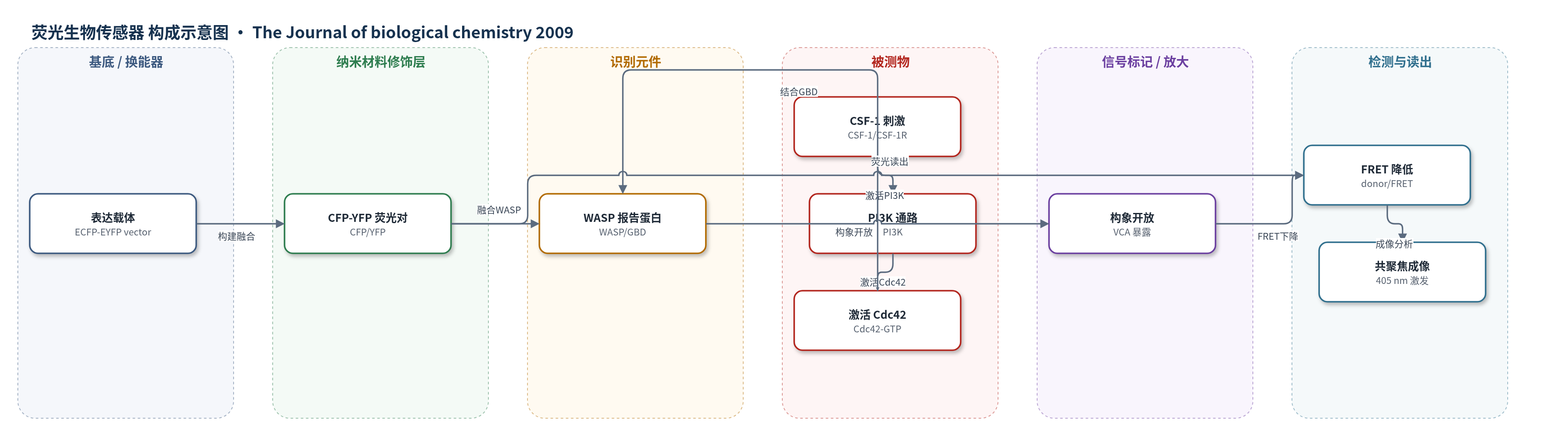
点击图片查看大图 · 依据论文自动绘制
传感器类型
荧光生物传感器
检测对象
WASP 激活状态(WASP activation)、CSF-1 诱导的 WASP 激活(CSF-1-induced WASP activation);样品基质:RAW/LR5 小鼠单核/巨噬细胞、HEK293/COS 细胞(活细胞或固定细胞)
检测原理
CSF-1 与巨噬细胞表面 CSF-1R 结合后,经 PI3K 通路激活 Cdc42,使其以 GTP 结合态(Cdc42-GTP)与 WASPbs 的 GBD 结合。该结合解除 WASP 的自抑制,使 VCA 区域暴露并促进 Arp2/3 相关肌动蛋白重塑。构象开放导致 CFP 与 YFP 距离增大,分子内 FRET 效率下降。用 405 nm 激光激发 CFP,检测 445–595 nm 发射光谱,以 donor/FRET 比值或 YFP 峰强度变化读出 WASP 激活程度;CSF-1 刺激增强时激活态比例增加,FRET 下降更明显。原文报告非激活态 FRET 效率约 19%,激活态约 5%,对应染料距离约 6.4 和 8.2 nm。该传感器不依赖酶促或核酸放大,而是直接以蛋白构象变化作为荧光信号。
检测灵敏度
原文未报告 LOD、线性范围、灵敏度斜率或相关系数。
效应效果
WASPbs 与构象敏感抗体(CSA)检测内源 WASP 的动力学一致:CSF-1 刺激后 30 s 出现显著激活,随后富集于细胞突起。非激活态 FRET 效率约 19%,激活态约 5%,对应染料距离 6.4 和 8.2 nm。wiskostatin 抑制 Cdc42Q61L 诱导的 FRET 下降,证明信号反映构象变化。Cdc42N17 降低基础激活并阻断 CSF-1 诱导激活;shRNA 使 Cdc42 降低 >70% 后激活消失。wortmannin/LY294002 阻断 PI3K 后激活消失,Cdc42V12 可绕过 PI3K。PP2、SU6656 和 Y291F 不影响激活,H246D 消除激活。作者认为该传感器可直接在活细胞内解析 CSF-1R 下游 WASP 调控,适用于巨噬细胞迁移与肿瘤进展研究。
传感器的构成
- 表达载体:ECFP-EYFP-biosensor vector,用于插入人 WASP 并构建 CFP-YFP 融合生物传感器
- 供体荧光标记:CFP(cyan fluorescent protein),融合于 WASP N 端,作为 FRET 供体
- 受体荧光标记:YFP(yellow fluorescent protein),融合于 WASP C 端,作为 FRET 受体
- 识别/报告元件:全长 WASP(Wiskott-Aldrich syndrome protein),其 GBD(GTPase-binding domain)结合激活 Cdc42 并发生构象变化
- 机制突变层:Y291F、H246D、L270P 等点突变,用于区分酪氨酸磷酸化、Cdc42 结合与开放构象
- 细胞表达基质:RAW/LR5 巨噬细胞、HEK293 或 COS 细胞,用于转染表达并受 CSF-1 刺激
中文摘要
本文报道了一种基于分子内荧光共振能量转移(FRET)的 Wiskott-Aldrich 综合征蛋白(WASP)生物传感器(WASPbs),用于在活细胞内实时监测 WASP 的激活状态。该传感器将青色荧光蛋白(CFP)和黄色荧光蛋白(YFP)分别融合于全长 WASP 的 N 端和 C 端;WASP 处于自抑制闭合构象时 CFP-YFP 距离较近,FRET 信号较强;当 WASP 被激活并发生构象开放时,二者距离增大,FRET 信号下降。将 WASPbs 转染巨噬细胞后,用集落刺激因子-1(CSF-1)刺激,30 s 内即可在全细胞范围检测到 WASP 激活,随后激活信号富集于细胞突起区域。采用构象敏感抗体检测内源 WASP 得到相似动力学。进一步机制研究显示,CSF-1 诱导的 WASP 激活完全依赖 Cdc42,并依赖磷脂酰肌醇 3-激酶(PI3K);而 Src 家族激酶抑制剂 PP2 或 SU6656 以及 WASPbs 主要酪氨酸磷酸化位点突变(Y291F)均不能降低 CSF-1 诱导的 WASP 激活。结果表明,CSF-1R 下游 WASP 激活受 PI3K 和 Cdc42 调控,体内未见 WASP 酪氨酸磷酸化促进其激活的证据。
英文摘要
A role for Wiskott-Aldrich syndrome protein (WASP) in chemotaxis to various agents has been demonstrated in monocyte-derived cell types. Although WASP has been shown to be activated by multiple mechanisms in vitro, it is unclear how WASP is regulated in vivo. A WASP biosensor (WASPbs), which uses intramolecular fluorescence resonance energy transfer to report WASP activation in vivo, was constructed, and following transfection of macrophages, activation of WASPbs upon treatment with colony-stimulating factor-1 (CSF-1) was detected globally as early as 30 s and remained localized to protrusive regions at later time points. Similar results were obtained when endogenous WASP activation was determined using conformation-sensitive antibodies. In vivo CSF-1-induced WASP activation was fully Cdc42-dependent. Activation of WASP in response to treatment with CSF-1 was also shown to be phosphatidylinositol 3-kinase-dependent. However, treatment with the Src family kinase inhibitors PP2 or SU6656 or disruption of the major tyrosine phosphorylation site of WASPbs (Y291F mutation) did not reduce the level of CSF-1-induced WASP activation. Our results indicate that WASP activation downstream of CSF-1R is phosphatidylinositol 3-kinase- and Cdc42-dependent consistent with an involvement of these molecules in macrophage migration. However, although tyrosine phosphorylation of WASP has been proposed to stimulate WASP activity, we found no evidence to indicate that this occurs in vivo.