组成图示
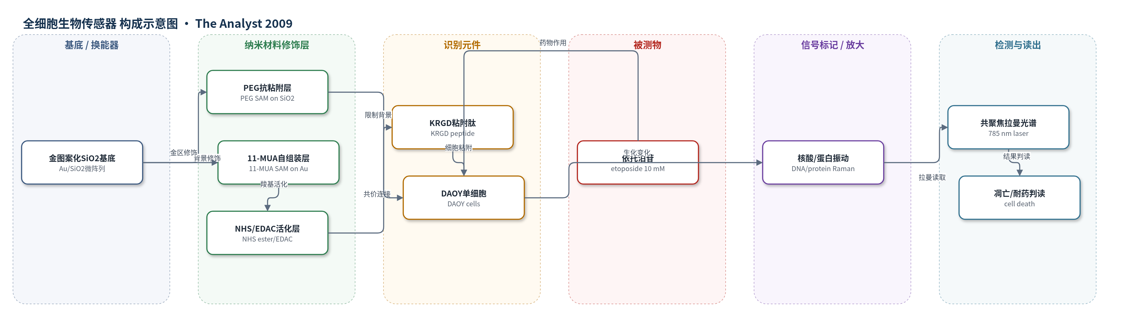
点击图片查看大图 · 依据论文自动绘制
传感器类型
全细胞生物传感器
检测对象
依托泊苷(etoposide)诱导的细胞凋亡(apoptosis)与细胞死亡(cell death);样品基质:Opti-MEM细胞培养液中的图案化DAOY单细胞阵列
检测原理
该传感器以图案化DAOY细胞为全细胞识别元件。依托泊苷进入细胞后抑制拓扑异构酶IIα,在DNA复制过程中造成DNA链断裂,触发凋亡。随着凋亡发生,细胞内核酸、蛋白质等生物分子含量和结构发生变化,细胞出现膜起泡、形态粗糙和细胞质量减少。共聚焦拉曼光谱基于分子振动的非弹性散射,无需化学标记即可检测核酸(782/788 cm-1)、苯丙氨酸(1005 cm-1)和蛋白/核酸C-H变形(1342 cm-1)等生物标志峰。药物暴露后,凋亡细胞的关键拉曼峰强度在24–36 h后显著下降,整体下降30–50%甚至超过50%;耐药细胞谱图基本保持。系统通过单细胞峰强度随时间变化判断细胞死亡或耐药,无外加化学放大,依赖原位长时间监测。
检测灵敏度
未报告LOD、线性范围、灵敏度斜率或相关系数。
效应效果
该平台在24个图案化DAOY单细胞(3组×8个)上实现48 h实时监测。10 mM依托泊苷处理后,87.5%细胞在36–48 h出现DNA/蛋白拉曼峰显著下降,12.5%细胞关键峰几乎不变,显示耐药亚群。图案化细胞群体与汇合培养细胞的PI流式细胞术存活曲线趋势一致,1342 cm-1拉曼峰与PI荧光定量相关;TUNEL检测显示药物组DNA断裂荧光显著升高,与拉曼结果一致。作者认为该方法非破坏、可重复监测单细胞,能识别低至10%的亚群(24细胞样本下超过90%概率),适用于药物筛选、毒素检测和生物威胁检测。
传感器的构成
- 基底/换能器:金图案化二氧化硅基底(Au/SiO2),提供20 μm×20 μm金方块微阵列和支撑
- 抗细胞粘附修饰层:聚乙二醇硅烷(PEG)在SiO2背景形成自组装单层(PEG SAM),排斥细胞
- 细胞粘附修饰层:11-巯基十一烷酸(11-MUA)在金方块上形成自组装单层(SAM),提供羧基
- 活化连接层:N-羟基琥珀酰亚胺(NHS)与EDAC活化11-MUA羧基形成NHS酯,用于共价连接肽
- 识别/粘附元件:赖氨酸-精氨酸-甘氨酸-天冬氨酸肽(KRGD)连接NHS酯,通过αvβ3整合素增强DAOY细胞粘附
- 传感元件:人小脑母细胞瘤DAOY细胞(DAOY cells)作为全细胞识别元件,响应依托泊苷
- 信号读出:共聚焦拉曼光谱(confocal Raman spectroscopy),无需化学标记,读取核酸/蛋白振动峰
中文摘要
本文报道了一种基于细胞的生物传感器应用,将图案化单细胞阵列与共聚焦拉曼光谱相结合,用于实时观察单个细胞对药物的时间依赖性反应。该图案化单细胞平台可方便地定位单个细胞并持续寻址,以非破坏、定量的方式表征其生化组成变化,从而保留离散细胞行为和细胞间差异。研究中,人小脑母细胞瘤DAOY细胞暴露于常用化疗药物依托泊苷,并在48小时内记录图案化细胞的拉曼光谱。结果显示,87.5%的监测细胞在药物暴露48小时后与DNA和蛋白质相关的拉曼峰显著下降,对应细胞死亡;其余12.5%细胞的关键拉曼生物标志物几乎无下降,表明其具有耐药性。此外,图案化细胞群体对依托泊苷的反应与汇合细胞培养物非常相似,并经流式细胞术证实。最后,采用TUNEL检测评估依托泊苷诱导的DNA断裂和凋亡,结果与拉曼光谱分析一致。该生物传感器-拉曼平台为高通量评估细胞对化学和生物试剂的反应提供了快速简便方法,可潜在用于药物发现、毒素检测和生物威胁检测。
英文摘要
We report on a cell-based biosensor application that utilizes patterned single-cell arrays combined with confocal Raman spectroscopy to observe the time-dependent drug response of individual cells in real time. The patterned single-cell platform enables individual cells to be easily located and continuously addressable for Raman spectroscopy characterization of biochemical compositional changes in a non-destructive, quantitative manner so that discrete cellular behavior and cell-to-cell variations are preserved. In this study, human medulloblastoma (DAOY) cells were exposed to the common chemotherapeutic agent etoposide, and Raman spectra from patterned cells were recorded over 48 hours. It was found that 87.5% of the cells monitored exhibited a sharp decrease in DNA and protein associated peaks 48 hours after drug exposure, corresponding to cell death. The remaining 12.5% of the cells showed little to no reduction in key Raman biomarkers, indicating their drug resistance. Furthermore, the patterned cell population showed a very similar response to etoposide as confluent cell cultures, as confirmed by flow cytometry. Finally, patterned cells were assessed with TUNEL assay for apoptosis due to DNA fragmentation after etoposide exposure. The results agree well with those from the Raman spectroscopy analysis. This combined biosensor-Raman platform provides a quick, simple way to assess cell responses to chemical and biological agents with high throughput and can be potentially used for a wide variety of biomedical applications such as pharmaceutical drug discovery, toxin tests, and biothreat detection.