组成图示
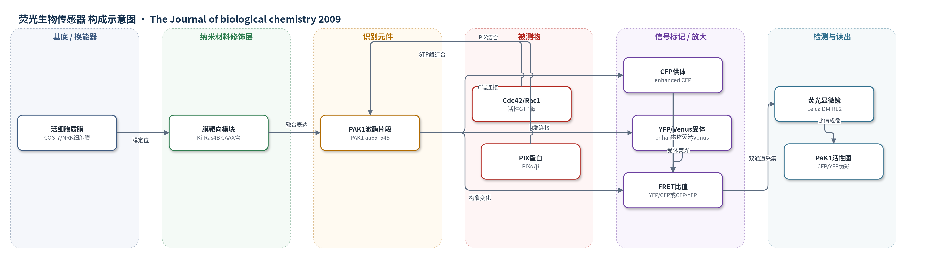
点击图片查看大图 · 依据论文自动绘制
传感器类型
荧光生物传感器
检测对象
PAK1激酶活性/构象状态(p21-activated kinase 1, PAK1),样品基质为活细胞(COS-7、NRK、HEK-HT、RPE1等)
检测原理
该传感器为FRET构象探针:非活性PAK1以交叉抑制二聚体呈闭合构象,N端YFP/Venus与C端CFP距离较近,CFP激发后能量转移至YFP,YFP/CFP比值较高。当活性Cdc42或Rac1结合PAK1的CRIB基序,或PAK1被招募至质膜并与PIX相互作用时,PAK1发生构象重排、二聚体解离,YFP与CFP距离增大,FRET效率下降,YFP/CFP降低而CFP/YFP升高。膜靶向Pakabix使PAK1在质膜局部富集,形成半开放中间态,Ser199/204磷酸化促进该状态,但对GTP酶刺激更敏感;完全开放态伴随Thr423磷酸化。信号通过双发射荧光比值读出,无化学放大,但膜局部富集和分子间反式磷酸化增强激活持续性。
检测灵敏度
R^2 = 0.62(Cdc42V12);R^2 = 0.49(Rac1V12)
效应效果
Pakabi的FRET变化与磷酸化抗体检测一致,并随Cdc42V12表达量剂量依赖下降;CRIB突变体不响应Cdc42V12,说明特异性。膜靶向Pakabix较胞质Pakabi的FRET仅降低6%(p<0.0001),加入Cdc42V12后进一步降低26%,表明膜招募诱导半开放态且对GTP酶高敏感。CRIB和R193A分别阻断GTP酶与PIX结合,双突变使突起局部PAK1激活降至K299R水平。siRNA耗竭PAK1约80%后抑制HEK-HT细胞铺展,PAK2耗竭无此效应;YFP-PID抑制PAK1活性也降低细胞面积。
传感器的构成
- 荧光供体:增强型CFP(enhanced CFP,K26R/Y66W/D129G/N146I/M153T/V163A/N164H/S175G),位于PAK1 C端,提供FRET供体荧光
- 识别/报告元件:人源PAK1激酶C端片段(PAK1 aa65–545),含CRIB和PID相关区域,感知Cdc42/Rac1结合并发生构象变化
- 荧光受体:增强型YFP或Venus(enhanced YFP/Venus,T65G/V68L/S72A/M153T/V163A/S175G/T203Y),位于PAK1 N端,作为FRET受体
- 连接肽:Leu-Asp-Thr-Met与Cys-Gly-Arg间隔序列,连接荧光蛋白与PAK1片段
- 膜靶向模块:Pakabix C端Gly-Arg-Ser-Arg间隔及Ki-Ras4B C端区(aa169–188,含CAAX盒),将传感器靶向质膜
- 突变对照元件:CRIB突变(aa81–87 ASAASAA)阻断GTP酶结合,R193A阻断PIX结合,K299R激酶失活,L107F组成激活
- 表达载体:pRaichu-Ras家族/pCAGGS真核表达载体,用于COS-7、NRK等细胞转染表达
中文摘要
p21激活激酶1(PAK1)是Cdc42和Rac1 GTP酶的关键效应子,通过调控肌动蛋白细胞骨架和黏附动态控制细胞突起与运动,其失调与人类癌症相关。本文报道了一种基于荧光共振能量转移(FRET)的PAK1构象生物传感器,用于在活细胞中直接观察PAK1激活的动态过程。该传感器由YFP、PAK1 C端片段和CFP融合组成,PAK1激活导致构象打开、二聚体解离,使供体CFP与受体YFP距离增加,FRET比值下降。利用膜靶向Pakabix,作者观察到COS-7细胞铺展和正常大鼠肾细胞(NRK)迁移过程中,PAK1在新形成突起和前缘板状伪足处被动态激活。研究还发现,PAK1招募至质膜后形成半开放中间态,选择性磷酸化N端调控区Ser199/204但不磷酸化催化环Thr423;该中间态对Cdc42/Rac1刺激高度敏感;PIX蛋白以GTP酶非依赖方式促进突起膜处PAK1激活;膜上PAK1分子间反式磷酸化可能参与激活。该工作提出了PAK1在细胞突起处多步骤、多信号整合的调控模型。
英文摘要
The p21-activated kinase (PAK) 1 kinase, an effector of the Cdc42 and Rac1 GTPases, regulates cell protrusions and motility by controlling actin and adhesion dynamics. Its deregulation has been linked to human cancer. We show here that activation of PAK1 is necessary for protrusive activity during cell spreading. To investigate PAK1 activation dynamics at live protrusions, we developed a conformational biosensor, based on fluorescence resonance energy transfer. This novel PAK1 biosensor allowed the spatiotemporal visualization of PAK1 activation during spreading of COS-7 cells and during motility of normal rat kidney cells. By using this imaging approach in COS-7 cells, the following new insights on PAK1 regulation were unveiled. First, PAK1 acquires an intermediate semi-open conformational state upon recruitment to the plasma membrane. This semi-open PAK1 species is selectively autophosphorylated on serines in the N-terminal regulatory region but not on the critical threonine 423 in the catalytic site. Second, this intermediate PAK1 state is hypersensitive to stimulation by Cdc42 and Rac1. Third, interaction with PIX proteins contributes to PAK1 stimulation at membrane protrusions, in a GTPase-independent way. Finally, trans-phosphorylation events occur between PAK1 molecules at the membrane possibly playing a relevant role for its activation. This study leads to a model for the complex and accurate regulation of PAK1 kinase in vivo at cell protrusions.