组成图示
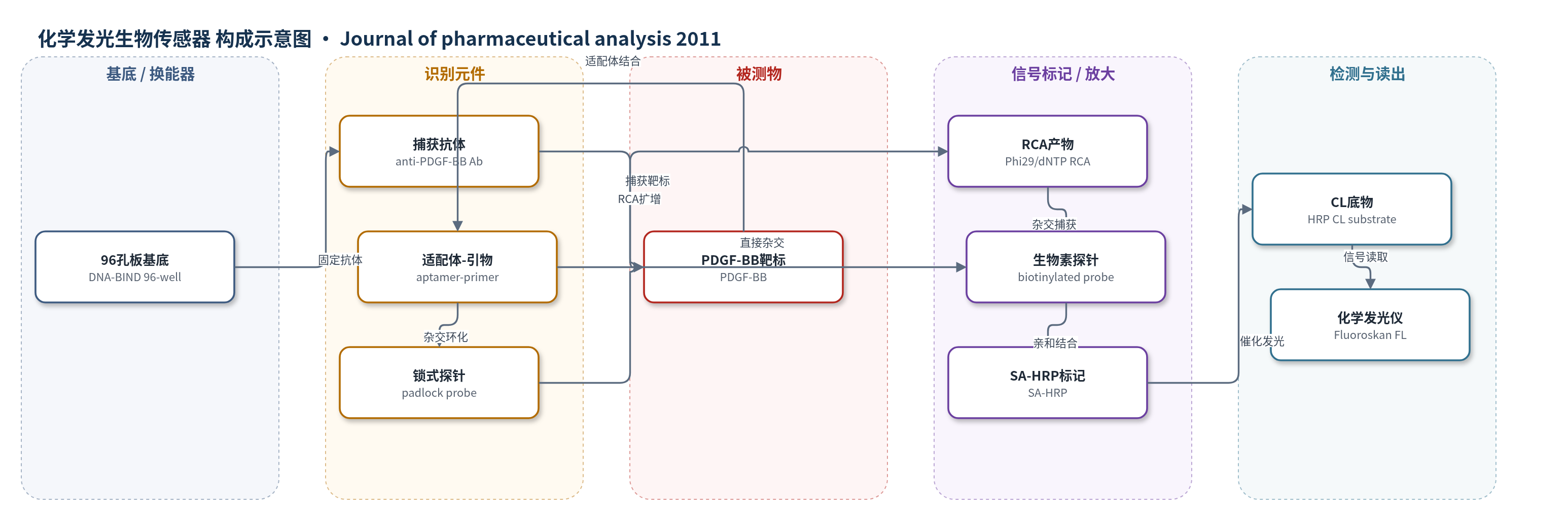
点击图片查看大图 · 依据论文自动绘制
传感器类型
化学发光生物传感器
检测对象
血小板源性生长因子B链(PDGF-BB,platelet-derived growth factor B-chain);样品基质:PBS 缓冲液/蛋白稀释液(未报告临床样品)
检测原理
固定于96孔板的抗PDGF-BB抗体捕获样品中的PDGF-BB,形成免疫夹心;与靶标结合的适配体–引物序列中,适配体部分完成识别,3'端引物与padlock probe两端杂交,经大肠杆菌DNA连接酶环化形成RCA模板。Phi29 DNA聚合酶在dNTP存在下以该模板进行滚环扩增,生成含多个串联重复序列的单链DNA产物。生物素化探针与RCA产物杂交,随后SA-HRP通过生物素–链霉亲和素高亲和结合被捕获。加入HRP化学发光底物后,HRP催化底物产生化学发光,发光强度随PDGF-BB浓度增加而增强;RCA使单个靶标事件转化为多个生物素位点,实现约1000倍灵敏度提升。
检测灵敏度
LOD: 10 fM;线性范围: 10 fM–1 nM;lgI=0.836 lgC+0.928;R^2 = 0.997;无RCA线性范围: 10 pM–10 nM;lgI=0.879 lgC-0.446;R^2 = 0.998
效应效果
该方法在优化条件下对PDGF-BB具有fM级灵敏度,RCA使灵敏度较无RCA夹心法提高1000倍。选择性方面,1 nM非靶蛋白IgG、IgA、IgM、干扰素和凝血酶相对于100 pM PDGF-BB产生的CL信号较低,表明信号主要由适配体–靶标结合触发,作者称可区分10倍浓度其他蛋白。文中未报告稳定性、重现性RSD或实际样品加标回收率。与已报道适配体法相比,本方法LOD 10 fM,优于或可比荧光、电化学、比色、毛细管电泳等PDGF-BB检测。作者认为其无需光源、建立简便,可用作PDGF-BB临床诊断工具,并易于扩展至IgE、凝血酶等其他疾病相关生物标志物。
传感器的构成
- 基底/载体:DNA-BIND 96-well plate(N-oxysuccinimide 表面,用于抗体固定与样品分离)
- 捕获识别层:goat anti-human PDGF-BB antibody(固定于孔板,捕获 PDGF-BB)
- 封闭层:5% BSA blocking buffer(封闭非特异结合位点)
- 识别/信号启动层:aptamer–primer(适配体识别 PDGF-BB,3'端引物启动 RCA)
- 环化模板:padlock probe(与 aptamer–primer 两端杂交,经 E. coli DNA ligase 环化)
- 扩增元件:Phi29 DNA polymerase 与 dNTPs(催化 RCA 生成串联重复单链 DNA)
- 信号探针:biotinylated probe(杂交 RCA 产物,携带生物素)
- 信号放大标记:SA–HRP(链霉亲和素–辣根过氧化物酶,结合生物素并催化 CL 底物发光)
- 读出层:commercial CL HRP substrate 与 Fluoroskan Ascent FL(HRP 催化底物产生化学发光,仪器读取)
中文摘要
本文报道了一种稳健、选择性且高灵敏的化学发光(CL)蛋白检测平台。该方法以滚环扩增(RCA)作为信号增强策略,以96孔板作为固定与分离载体。典型流程中,先将抗体固定于96孔板表面,再与目标蛋白及适配体–引物序列形成夹心复合物;该适配体–引物序列随后作为RCA引物。基于此设计,可在孔板表面捕获大量生物素化探针及链霉亲和素–辣根过氧化物酶(SA–HRP),从而放大化学发光信号。结果表明,该生物传感器检测限达10 fM,建立简便、易于使用且无需外部光源。因此,该技术有望拓展基于DNA的生物传感器在临床疾病相关蛋白生物标志物检测中的应用,兼具高灵敏度与高选择性。
英文摘要
A robust, selective and highly sensitive chemiluminescent (CL) platform for protein assay was presented in this paper. This novel CL approach utilized rolling circle amplification (RCA) as a signal enhancement technique and the 96-well plate as the immobilization and separation carrier. Typically, the antibody immobilized on the surface of 96-well plate was sandwiched with the protein target and the aptamer-primer sequence. This aptamer-primer sequence was then employed as the primer of RCA. Based on this design, a number of the biotinylated probes and streptavidin-horseradish peroxidase (SA-HRP) were captured on the plate, and the CL signal was amplified. In summary, our results demonstrated a robust biosensor with a detection limit of 10 fM that is easy to be established and utilized, and devoid of light source. Therefore, this new technique will broaden the perspective for future development of DNA-based biosensors for the detection of other protein biomarkers related to clinical diseases, by taking advantages of high sensitivity and selectivity.