组成图示
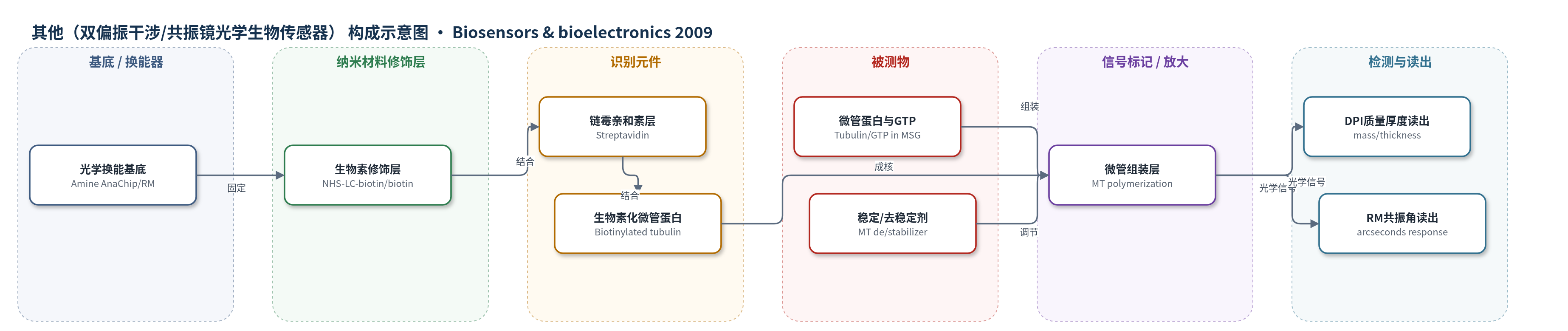
点击图片查看大图 · 依据论文自动绘制
传感器类型
其他(双偏振干涉/共振镜光学生物传感器)
检测对象
微管扰动剂(MT-perturbing agents:paclitaxel 紫杉醇、epothilone B、colchicine 秋水仙碱、dolastatin 10)及微管蛋白组装(tubulin assembly/microtubules, MTs);样品基质:含GTP的1 M MSG缓冲液中的牛脑微管蛋白溶液。
检测原理
传感器表面通过生物素—链霉亲和素—生物素化微管蛋白构建微管成核层。将含GTP的微管蛋白加入1 M MSG缓冲液后,微管蛋白二聚体在表面结合并纵向组装成原丝,随后约13条原丝横向连接形成中空微管。该过程使传感层质量与厚度增加、密度下降。DPI同时测量TM和TE偏振干涉,解算有效折射率、厚度、质量和密度;RM通过高折射率共振层检测表面折射率变化引起的共振角位移(arcseconds)。微管稳定剂促进组装并提高初始速率斜率,去稳定剂抑制组装并降低斜率,因此可通过初始15 s响应斜率区分稳定剂与去稳定剂。
检测灵敏度
未报告LOD、线性范围与R^2;初始组装速率斜率: 1.50 ± 0.27 arcseconds/s(paclitaxel)、1.04 ± 0.13 arcseconds/s(epothilone B)、0.37 ± 0.11 arcseconds/s(GTP对照)、0.05 ± 0.01 arcseconds/s(colchicine)、0.27 ± 0.08 arcseconds/s(dolastatin 10);DPI厚度生长速率: 0.236 nm/min、0.436 nm/min。
效应效果
DPI可分辨质量、厚度与密度变化,证实表面微管形成;RM比色皿样品量小、可重复使用,微管蛋白组装在约20个循环内可重复,但室温过夜或4 ℃保存后活性丧失。温度依赖性与浊度法结果一致。与仅含GTP对照相比,paclitaxel、epothilone B、colchicine和dolastatin 10的初始速率斜率成对P值分别为0.035、0.005、0.033和0.838(n=3),表明可区分稳定剂与去稳定剂。colchicine预孵育可完全抑制结合,paclitaxel使响应更稳定。未报告RSD与实际样品回收率。作者认为该方法可用于高通量筛选微管扰动剂。
传感器的构成
- 基底/换能器:Amine AnaChip 双偏振干涉波导芯片或 biotin cuvette 共振镜比色皿,提供光学换能表面与固定位点
- 表面修饰层:NHS-LC-biotin 生物素化氨基芯片(DPI)或 biotin cuvette 生物素化表面(RM),形成生物素捕获层
- 捕获层:Streptavidin 链霉亲和素,特异性结合生物素化微管蛋白
- 识别/成核层:Biotinylated tubulin 生物素化牛脑微管蛋白,作为微管组装起始模板
- 信号产生层:Tubulin 微管蛋白与 GTP 在 1 M MSG 缓冲液中组装成微管(MT),引起质量/厚度/密度变化
- 读出系统:DPI 双偏振干涉仪或 RM 共振镜,检测折射率、厚度、质量或共振角变化
中文摘要
本研究采用双偏振干涉(DPI)和共振镜(RM)两种表面光学生物传感器方法,表征微管(MTs)在生物传感器表面上的组装与动态。微管在细胞分裂中起关键作用,也是多种抗癌药物的靶点。DPI结果显示,在链霉亲和素—生物素化微管蛋白表面上加入微管蛋白后,传感层质量和厚度持续增加,同时密度下降,表明微管在表面垂直生长并形成中空结构。初始厚度增长速率为0.236 nm/min,提示原丝先纵向延长;随后原丝横向连接形成微管,生长速率增至0.436 nm/min。RM表面也观察到微管蛋白结合引起的连续质量增加。微管蛋白结合具有温度依赖性:15至37 ℃升温时,微管稳定剂使绝对响应增强,而去稳定剂抑制结合。在微管稳定剂存在下,初始组装速率斜率显著高于仅含GTP对照(1.50±0.27和1.04±0.13 arcseconds/s,对照0.37±0.11 arcseconds/s);秋水仙碱和dolastatin 10等去稳定剂使斜率降至0.05±0.01和0.27±0.08 arcseconds/s。结果表明,表面光学传感器可区分微管稳定剂与去稳定剂,并为筛选微管扰动剂提供新途径。
英文摘要
Dual polarization interferometry (DPI) and resonant mirror (RM) methods were used to characterize the growth of microtubules (MTs) on biosensor surfaces. The structure and dynamics of MTs play an important role in cell division and are a target for many anti-cancer drugs. Evidence from DPI demonstrated the growth of MTs on streptavidin-biotinylated-tubulin surfaces from the increase in mass and thickness, with a simultaneous decrease in density. The initial increase in thickness of 0.236 nm/min suggested the elongation of protofilaments before they join laterally to form the MT, where the rate of growth increased to 0.436 nm/min. Continuous mass increases were also observed when tubulin was added to a similar underlying RM surface. Tubulin binding to these surfaces was also temperature dependent, increasing the absolute response with MT stabilizers, while inhibiting binding with destabilizers when temperature was changed from 15 to 37 degrees C. Finally, the initial rates of tubulin assembly (mean+/-SD, n=3) with MT-stabilizer agents were significantly higher at 1.50+/-0.27 and 1.04+/-0.13 arcseconds/s, respectively, compared to 0.37+/-0.11 arcseconds/s for tubulin containing GTP only. In the presence of the MT destabilizers, colchicine and dolastatin 10, the slopes of initial rates were lower than in their absence at 0.05+/-0.01 and 0.27+/-0.08 arcseconds/s, respectively. This provides evidence for the ability of surface-based optical sensors to distinguish between MT stabilizers and destabilizers, while also paving the path to develop other methods to screen for MT-perturbing agents using the same underlying surface engineering.