组成图示
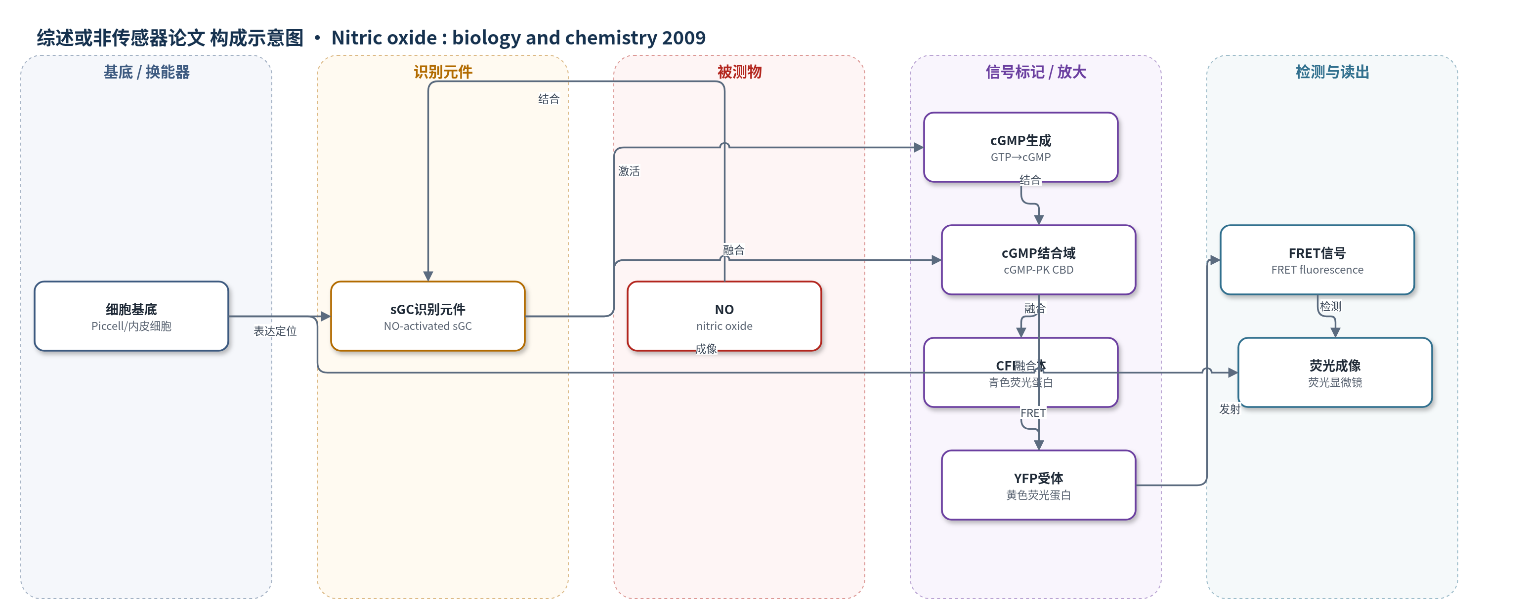
点击图片查看大图 · 依据论文自动绘制
传感器类型
综述或非传感器论文
检测对象
一氧化氮(NO, nitric oxide);样品基质:培养血管内皮细胞、神经元、脑组织、血管等细胞/组织中的生理或病理 NO 浓度
检测原理
该文综述的 NO 生物传感器以 NO 激活的可溶性鸟苷酸环化酶(sGC)为识别元件。NO 结合 sGC 后激活酶活性,催化 GTP 生成 cGMP;cGMP 与融合在 sGC 亚基上的 cGMP 依赖性蛋白激酶的 cGMP 结合域(cGMP-PK CBD)结合,引起青色荧光蛋白(CFP)与黄色荧光蛋白(YFP)之间的荧光共振能量转移(FRET)变化。FRET 信号强度随 cGMP 浓度及 NO 暴露水平改变,从而将 NO 浓度转换为可定量荧光输出。文中还讨论以组织内源 sGC/cGMP 作为 NO 生物传感器:NO 升高使 cGMP 积累,cGMP 水平或免疫染色可反推 NO 浓度。该策略利用生理受体选择性,避免电极对多巴胺、抗坏血酸等物质的交叉反应。
检测灵敏度
LOD: 0.1 nM;LOD: 20 pM;NO detection limit: 17 nM after 5 min exposure
效应效果
直接电化学 NO 电极结果差异极大,不同组织报道值跨约 10^6 个数量级,主要受亚硝酸根、多巴胺、5-羟色胺、酪氨酸、抗坏血酸等交叉反应影响;NO/多巴胺选择性 18:1、NO/抗坏血酸 750:1,脑内 500 μM 抗坏血酸变化可被误读为 330–660 nM NO。基于 sGC 的 NO 生物传感器选择性高,CO 需 100 μM 才产生可检测响应,NO 检出限 0.1 nM,Piccell 可达 20 pM;内皮细胞基础 NO 约 1 nM,ATP 光释放可产生约 100 pM 瞬时 NO,20 μm 处约 30 pM,神经元峰值约 100 pM。作者主张生理 NO 浓度约为 100 pM–5 nM,远低于早期 μM 级估计。
传感器的构成
- 细胞基底:培养血管内皮细胞或稳定表达传感器的细胞系 Piccell,用于承载 NO 生物传感器并模拟细胞间 NO 释放
- 识别元件:NO 激活的可溶性鸟苷酸环化酶(sGC),特异性结合 NO 并作为生理 NO 受体
- 信号放大元件:sGC 催化 GTP 生成 cGMP,将 NO 结合事件转化为第二信使浓度变化
- 荧光报告元件:cGMP 依赖性蛋白激酶的 cGMP 结合域(cGMP-PK CBD)与青色荧光蛋白(CFP)、黄色荧光蛋白(YFP)融合,cGMP 结合后产生 FRET 信号
- 读出元件:FRET 荧光变化,用于定量 NO 浓度
中文摘要
明确体内生理性一氧化氮(NO)浓度对于定量理解 NO 信号、开展贴近真实生理状态的 NO 实验以及判断疾病状态下 NO 是否异常至关重要。十年前,基于早期电极测量和 NO 对其鸟苷酸环化酶偶联受体效力的初步估计,约 1 μM 的 NO 浓度曾被认为是合理的。此后,许多在细胞和组织中用电极直接测定 NO 浓度的努力给出了差异极大且难以调和的结果。作为补充,多种替代方法的数据现已趋于一致,提供了更连贯的认识。这些方法包括对 NO 激活鸟苷酸环化酶的定量分析、基于细胞内 NO 合酶类型/活性/含量的计算机建模、利用新型生物传感器监测单个内皮细胞和神经元释放 NO,以及将鸟苷酸环化酶作为组织在多种刺激下的内源性 NO 生物传感器。所有这些独立证据均提示生理 NO 浓度范围约为 100 pM(或更低)至约 5 nM,比过去认为的低数个数量级。
英文摘要
Clarity about the nitric oxide (NO) concentrations existing physiologically is essential for developing a quantitative understanding of NO signalling, for performing experiments with NO that emulate reality, and for knowing whether or not NO concentrations become abnormal in disease states. A decade ago, a value of about 1 microM seemed reasonable based on early electrode measurements and a provisional estimate of the potency of NO for its guanylyl cyclase-coupled receptors, which mediate physiological NO signal transduction. Since then, numerous efforts to measure NO concentrations directly using electrodes in cells and tissues have yielded an irreconcilably large spread of values. In compensation, data from several alternative approaches have now converged to provide a more coherent picture. These approaches include the quantitative analysis of NO-activated guanylyl cyclase, computer modelling based on the type, activity and amount of NO synthase enzyme contained in cells, the use of novel biosensors to monitor NO release from single endothelial cells and neurones, and the use of guanylyl cyclase as an endogenous NO biosensor in tissue subjected to a variety of challenges. All these independent lines of evidence suggest the physiological NO concentration range to be 100 pM (or below) up to approximately 5 nM, orders of magnitude lower than was once thought.