组成图示
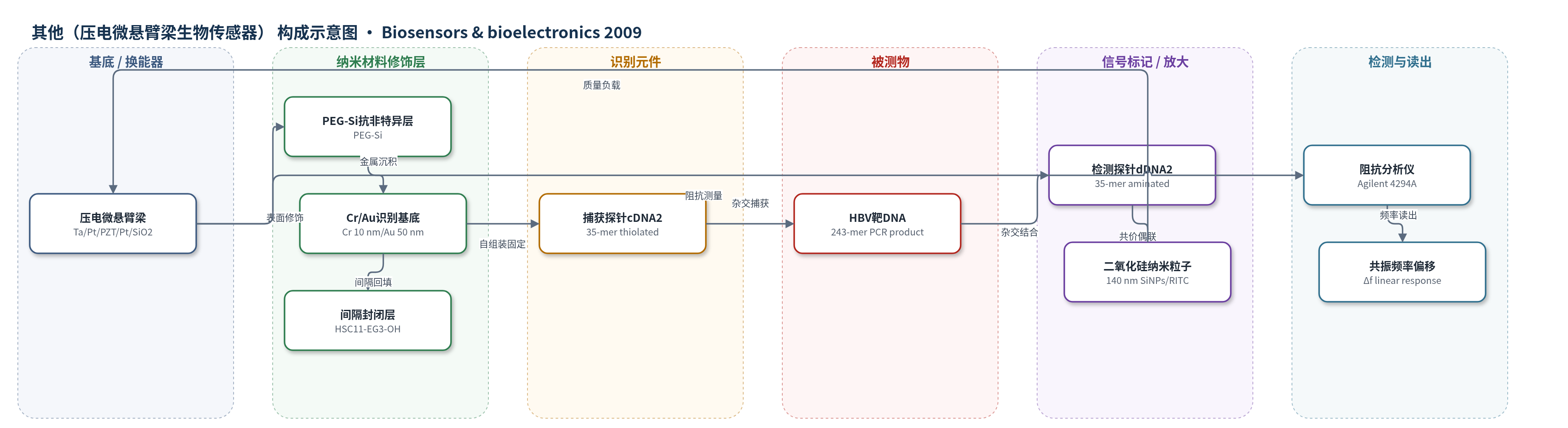
点击图片查看大图 · 依据论文自动绘制
传感器类型
其他(压电微悬臂梁生物传感器)
检测对象
HBV DNA(乙型肝炎病毒 DNA,243-mer precore/core 区 PCR 产物);样品基质:临床 HBV 阳性血清提取并经 nested PCR 扩增的 DNA,检测缓冲液为 TE buffer。
检测原理
微悬臂梁底面 Au 上固定硫醇化 cDNA2 捕获探针。HBV 靶 DNA 与 cDNA2 互补杂交,使悬臂梁表面质量增加;随后氨基化 dDNA2 与靶 DNA 另一端杂交,dDNA2 已通过 EDC/NHS 共价偶联到 140 nm 二氧化硅纳米粒子(SiNPs)上,形成夹心结构。SiNPs 提供大质量负载,实现信号放大。PZT 压电微悬臂梁在激励下共振,质量负载使共振频率下降;阻抗分析仪监测相位角/阻抗获得共振频率偏移。频率偏移与靶 DNA 浓度在 23.1 fM–2.3 nM 线性相关。作者认为线性主要来自质量增加,弹簧常数变化为次要因素。
检测灵敏度
LOD: ~2.3 fM(外推估计,原文未直接测量);线性范围: 23.1 fM–2.31 nM;共振频率偏移: 捕获步骤 -4 Hz 至 -187 Hz,纳米粒子增强步骤 -56 Hz 至 -413 Hz;灵敏度: 各浓度至少提高 2 倍,LOD 降低约 100 倍。
效应效果
该传感器对 25-mer 非互补 DNA 的负对照响应较小(DNA 结合步骤约 -7 Hz,纳米粒子增强步骤约 -24 Hz),表明杂交特异性较好;SEM 与荧光图像显示 2.3 pM HBV DNA 下 SiNPs 特异性结合于悬臂梁表面。每个浓度至少测试 10 根微悬臂梁,但未报告 RSD、稳定性或实际样品加标回收率。与无增强相比,纳米粒子增强使各浓度灵敏度至少提高 2 倍,LOD 降低约 100 倍,总体灵敏度提高约 2–3 个数量级。作者承认其灵敏度低于商业仪器(信号放大 <5000 copies/mL、靶标放大 <50 copies/mL),但系统更简单,无需复杂扩增或额外染色,具有用于 PCR-free 高灵敏 DNA 检测、病毒全序列检测、免疫分析、细胞/病毒计数及突变检测的潜力。
传感器的构成
- 基底/换能器:SiNx 支撑层上 Ta/Pt/PZT/Pt/SiO2 多层压电微悬臂梁,PZT 提供压电驱动与共振频率读出基础
- 抗非特异层:PEG-Si(2-[methoxy(polyethyleneoxy)propyl]trimethoxysilane)处理表面,减少 DNA 与生物分子非特异吸附
- 识别基底金属层:Cr/Au(10 nm/50 nm)电子束蒸发于悬臂梁底面,Cr 为粘附层,Au 提供 Au-S 结合位点
- 识别元件:硫醇化捕获探针 cDNA2(35-mer,5′-HS-T10 间隔)自组装于 Au 表面,与 HBV 靶 DNA 互补杂交
- 间隔封闭层:HSC11-EG3-OH spacer 回填,完善自组装单分子层,减少非特异结合并优化探针排列
- 信号标记物:氨基化检测探针 dDNA2(35-mer,5′-NH2)经 EDC/NHS 偶联到含 RITC 的 140 nm 二氧化硅纳米粒子(SiNPs)表面,形成 dDNA2-SiNPs
- 纳米粒子抗非特异层:SiNPs 表面 PEG(mw ~600)减少非特异结合,RITC 掺杂用于荧光验证
- 信号读出:阻抗分析仪(Agilent 4294A)监测相位角/阻抗,获得微悬臂梁共振频率偏移
中文摘要
本文报道了一种二氧化硅纳米粒子增强的动态微悬臂梁生物传感器,用于检测乙型肝炎病毒(HBV)DNA。以 HBV DNA precore/core 区的 243 核苷酸片段为靶标,设计了固定于微悬臂梁表面的捕获探针和偶联二氧化硅纳米粒子的检测探针。通过 DNA 杂交直接结合纳米粒子,优化了二氧化硅纳米粒子尺寸与微悬臂梁尺寸,并讨论了纳米粒子浓度与微悬臂梁共振频率偏移之间的定量关系,以验证质量负载与频率偏移的关联。利用 PCR 产物,在 23.1 fM 至 2.31 nM 范围内检测了 HBV 靶 DNA;无纳米粒子增强时可检测至皮摩尔级,使用纳米粒子信号放大后可检测至飞摩尔级。两种情况下,共振频率偏移均与 HBV 靶 DNA 浓度呈线性相关。作者认为该线性主要来自探针 DNA 与 HBV PCR 产物、以及 HBV PCR 产物与二氧化硅纳米粒子结合所导致的质量增加,尽管弹簧常数变化也可能影响微悬臂梁共振频率。
英文摘要
We report Hepatitis B Virus (HBV) DNA detection using a silica nanoparticle-enhanced dynamic microcantilever biosensor. A 243-mer nucleotide of HBV DNA precore/core region was used as the target DNA. For this assay, the capture probe on the microcantilever surface and the detection probe conjugated with silica nanoparticles were designed specifically for the target DNA. For efficient detection of the HBV target DNA using silica nanoparticle-enhanced DNA assay, the size of silica nanoparticles and the dimension of microcantilever were optimized by directly binding the silica nanoparticles through DNA hybridization. In addition, the correlation between the applied nanoparticle concentrations and the resonant frequency shifts of the microcantilever was discussed clearly to validate the quantitative relationship between mass loading and resonant frequency shift. HBV target DNAs of 23.1 fM to 2.31 nM which were obtained from the PCR product were detected using a silica nanoparticle-enhanced microcantilever. The HBV target DNA of 243-mer was detected up to the picomolar (pM) level without nanoparticle enhancement and up to the femtomolar (fM) level using a nanoparticle-based signal amplification process. In the above two cases, the resonant frequency shifts were found to be linearly correlated with the concentrations of HBV target DNAs. We believe that this linearity originated mainly from an increase in mass that resulted from binding between the probe DNA and HBV PCR product, and between HBV PCR product and silica nanoparticles for the signal enhancement, even though there is another potential factor such as the spring constant change that may have influenced on the resonant frequency of the microcantilever.