组成图示
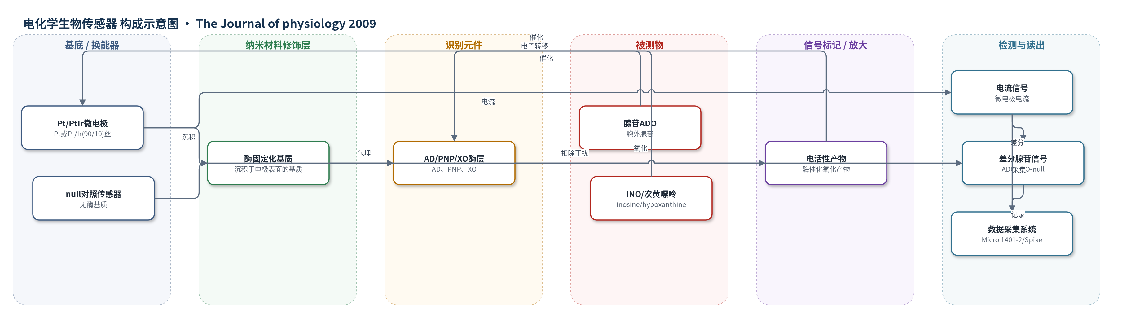
点击图片查看大图 · 依据论文自动绘制
传感器类型
电化学生物传感器
检测对象
腺苷(adenosine, ADO)、次黄嘌呤核苷(inosine, INO)、次黄嘌呤(hypoxanthine);样品基质:大鼠小脑切片分子层细胞外液(人工脑脊液 aCSF 灌注)
检测原理
胞外腺苷或次黄嘌呤核苷扩散至微电极表面,被固定化酶层催化:腺苷脱氨酶(AD)将腺苷转化为次黄嘌呤核苷,嘌呤核苷磷酸化酶(PNP)将次黄嘌呤核苷转化为次黄嘌呤,黄嘌呤氧化酶(XO)进一步氧化次黄嘌呤/黄嘌呤生成电活性产物并释放电子。电子在 Pt/PtIr 微电极上被检测,形成与嘌呤浓度相关的电流。传感器经筛选以降低 5-HT、去甲肾上腺素、多巴胺和抗坏血酸等干扰。ADO/INO 传感器与无酶 null 传感器差分,可区分腺苷及其代谢物。浓度越高,酶催化产物越多,电流越大;原文报告响应线性、上升时间小于 10 s。
检测灵敏度
检测水平: ~30 nM(原文:below the levels of detection (~30 nM))
效应效果
传感器经筛选后对 5-HT、去甲肾上腺素、多巴胺和抗坏血酸等电活性干扰物响应降低,并用 10 μM 腺苷/次黄嘌呤核苷校准、10 μM 5-HT 检查筛选。基础状态下 null 传感器无基线变化,ADO/INO 传感器仅检测到代谢物;P9–12 切片表面次黄嘌呤核苷约 0.25±0.03 μM,P21–28 约 1.6±0.3 μM。缺氧 10 min 后最大次黄嘌呤核苷 P9–12 为 3.5±0.4 μM、P21–28 为 5.5±0.7 μM,上升时间常数 9.1±0.4 min 对 1.5±0.3 min,衰减约 15 min。成熟片电刺激 4/4 可检测释放,未成熟片未检测到。
传感器的构成
- 基底/换能器电极:Pt 或 Pt/Ir(90/10) 丝,蚀刻至约50 μm,作为电化学换能器
- 酶固定化基质:沉积于电极表面的基质,包埋酶并构成传感膜
- 催化识别元件:腺苷脱氨酶(AD)、嘌呤核苷磷酸化酶(PNP)和黄嘌呤氧化酶(XO),将腺苷/次黄嘌呤核苷/次黄嘌呤转化为电活性产物
- 抗干扰筛选层:经筛选(screened)处理,降低5-HT、去甲肾上腺素、多巴胺和抗坏血酸等电活性干扰物响应
- 对照传感器:null sensor,仅含基质不含酶,用于扣除非特异电活性干扰
- 差分信号层:ADO/INO 传感器与 null 传感器差分,获得腺苷及其代谢物(次黄嘌呤核苷、次黄嘌呤)特异信号
中文摘要
腺苷是哺乳动物脑内重要的细胞外嘌呤信号分子,参与多种生理与病理过程,但其在脑发育过程中释放与清除如何变化仍不清楚。本研究将电生理记录与微电极生物传感器测量相结合,检测大鼠小脑出生后第9–12天、平行纤维–浦肯野细胞突触刚形成阶段的腺苷信号。结果显示,在基础状态和刺激序列中,A1受体介导的抑制性张力很小或缺失;抑制腺苷清除的药物对突触传递影响很小,提示基础状态下细胞外腺苷浓度变化很小。缺氧可诱导明显腺苷释放,但释放延迟且缓慢;分子层电刺激后未检测到腺苷释放。结论认为,该发育阶段虽然腺苷受体和清除机制已存在,但腺苷释放很少。
英文摘要
The purine adenosine is an extracellular signalling molecule involved in a large number of physiological and pathological conditions throughout the mammalian brain. However little is known about how adenosine release and its subsequent clearance change during brain development. We have combined electrophysiology and microelectrode biosensor measurements to investigate the properties of adenosine signalling at early stages of cerebellar development, when parallel fibre-Purkinje cell synapses have recently been formed (postnatal days 9-12). At this stage of development, we could detect little or no inhibitory A(1) receptor tone in basal conditions and during trains of stimuli. Addition of pharmacological agents, to inhibit adenosine clearance, had only minor effects on synaptic transmission suggesting that under basal conditions, the concentration of adenosine moving in and out of the extracellular space is small. Active adenosine release was stimulated with hypoxia and trains of electrical stimuli. Although hypoxia released significant concentrations of adenosine, the release was delayed and slow. No adenosine release could be detected following electrical stimulation in the molecular layer. In conclusion, at this stage of development, although adenosine receptors and the mechanisms of adenosine clearance are present there is very little adenosine release.