组成图示
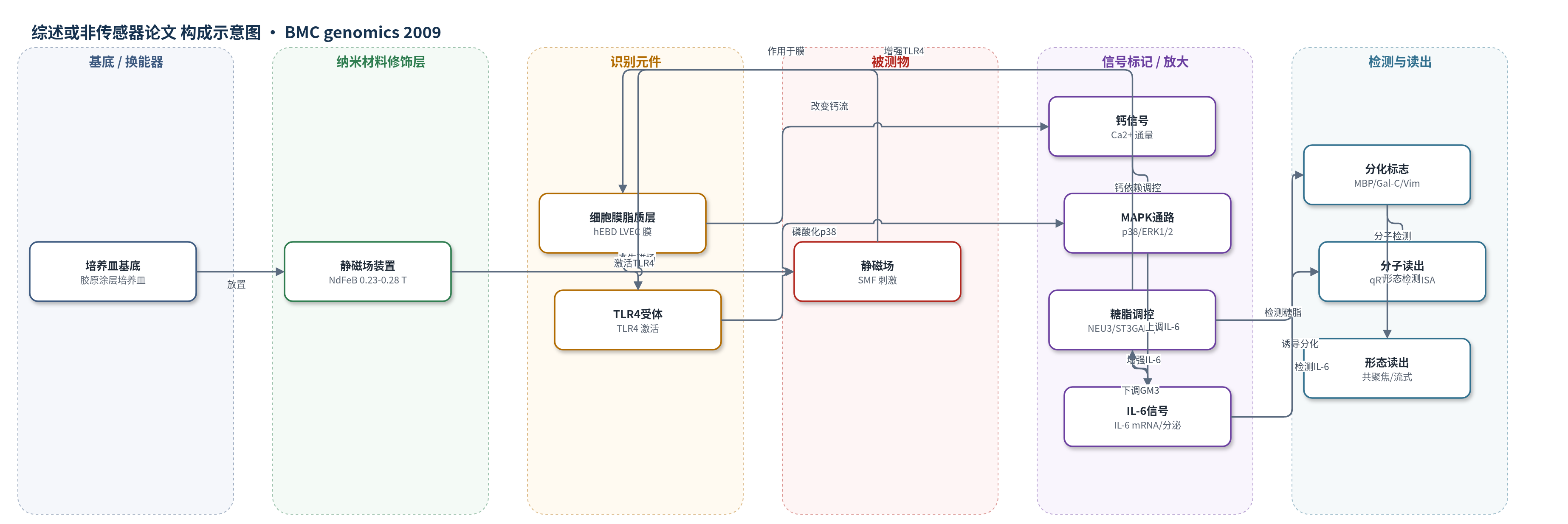
点击图片查看大图 · 依据论文自动绘制
传感器类型
综述或非传感器论文
检测对象
静磁场(SMF)暴露诱导的 IL-6、TLR4、NEU3、ST3GAL5、GM3/GD3 及少突胶质分化标志物;样品基质:hEBD LVEC 细胞培养体系
检测原理
静磁场(SMF)首先作用于 hEBD LVEC 细胞膜脂质双分子层,改变膜流动性/刚性并影响离子通道通量,尤其引起胞外/胞内 Ca2+ 分布变化。膜界面变化激活 TLR4,并通过 Ca2+ 依赖途径和 p38/ERK1/2 MAPK 磷酸化促进 IL-6 mRNA 转录与分泌。与此同时,SMF 独立上调 NEU3、下调 ST3GAL5,使神经节苷脂 GM3/GD3 降低;GM3 缺失解除其对 TLR4 和 IL-6 的抑制,形成前馈放大。长期暴露中 GM3 丢失又削弱 Ca2+ 依赖信号,导致 IL-6 响应衰减。最终信号表现为细胞形态改变及 MBP、Gal-C、Vim 等少突胶质前体标志物升高,而 BMP-2、GFAP 不升高。qRT-PCR、ELISA、流式细胞术、Western blot 和共聚焦显微镜用于读出上述分子与表型变化。
检测灵敏度
原文未报告。
效应效果
实验在 n≥3 重复中进行,多数 p<0.05。15 min SMF 仅 2 个基因显著变化且无 2 倍变化;24 h 有 379 个基因上调、549 个下调;5 d 有 85 个基因上调≥2 倍、94 个下调≥2 倍,恢复 1 d 后上调降至 47。IL-6 mRNA 2 h 升高并在 4、7、24 h 维持,分泌 IL-6 7 h 升高、48 h 最大、96 h 仍高于对照,6 d 回落。SMF 使 p38 磷酸化增加,3 d 增殖下降、9 d 消失,凋亡无变化;HEK AD293 无增殖改变。SMF 联合 4.0 ng/ml IL-6 使约 100% 细胞出现分化形态,MBP、Gal-C、Vim 升高,GFAP/NEF 不升高。作者认为可用于非侵入性神经分化调控。
传感器的构成
- 基底/换能器:胶原涂层组织培养塑料或玻璃底培养皿,承载 hEBD LVEC 细胞并支持成像
- 磁场刺激层:NdFeB 永磁体静磁场装置,提供 0.23–0.28 T 均匀磁场
- 识别/响应元件:hEBD LVEC 细胞膜脂质双分子层,作为静磁场响应的分子界面
- 信号标记物:SYBR Green、荧光抗体、HRP 二抗、钙离子试剂等,用于标记 mRNA、蛋白、糖脂和钙信号
- 读出层:qRT-PCR、ELISA、流式细胞仪、Western blot 和共聚焦显微镜,输出基因表达、蛋白分泌、糖脂水平和形态学信号
中文摘要
已有证据表明磁场可调节生命系统,但既往严格研究多集中于寻找分子水平生物传感器(如自由基离子对或细胞膜)或观察整体动物行为,导致分子效应如何转化为组织与个体水平响应存在空白。本研究通过人胚胎来源细胞的全 mRNA 芯片分析并结合软件通路分析,探讨 0.23–0.28 T 静磁场(SMF)对信号网络的影响。结果显示,SMF 暴露后共有九个信号网络发生响应;作者对与炎症细胞因子 IL-6 相关的网络进行了生化验证。短期(<24 h)IL-6 激活涉及 TLR4 的协同上调,以及 NEU3 和 ST3GAL5 的互补变化,使神经节苷脂 GM3 降低,从而增强 TLR4 与 IL-6 的激活。GM3 的丢失也为较长暴露时间后细胞响应减弱提供了可能机制。最终,SMF 诱导的细胞水平表现为形态改变及提示少突胶质前体分化的生化标志物。该研究提出了从脂质膜这一可能分子传感器到细胞分化等整体响应的框架,并揭示了神经节苷脂、IL-6 时间依赖调控与胚胎细胞命运之间的新关系。
英文摘要
BACKGROUND: Compelling evidence exists that magnetic fields modulate living systems. To date, however, rigorous studies have focused on identifying the molecular-level biosensor (e.g., radical ion pairs or membranes) or on the behavior of whole animals leaving a gap in understanding how molecular effects are translated into tissue-wide and organism-level responses. This study begins to bridge this gulf by investigating static magnetic fields (SMF) through global mRNA profiling in human embryonic cells coupled with software analysis to identify the affected signaling pathways.
RESULTS: Software analysis of gene expression in cells exposed to 0.23-0.28 T SMF showed that nine signaling networks responded to SMF; of these, detailed biochemical validation was performed for the network linked to the inflammatory cytokine IL-6. We found the short-term (<24 h) activation of IL-6 involved the coordinate up-regulation of toll-like receptor-4 (TLR4) with complementary changes to NEU3 and ST3GAL5 that reduced ganglioside GM3 in a manner that augmented the activation of TLR4 and IL-6. Loss of GM3 also provided a plausible mechanism for the attenuation of cellular responses to SMF that occurred over longer exposure periods. Finally, SMF-mediated responses were manifest at the cellular level as morphological changes and biochemical markers indicative of pre-oligodendrocyte differentiation.
CONCLUSION: This study provides a framework describing how magnetic exposure is transduced from a plausible molecular biosensor (lipid membranes) to cell-level responses that include differentiation toward neural lineages. In addition, SMF provided a stimulus that uncovered new relationships - that exist even in the absence of magnetic fields - between gangliosides, the time-dependent regulation of IL-6 signaling by these glycosphingolipids, and the fate of embryonic cells.