组成图示
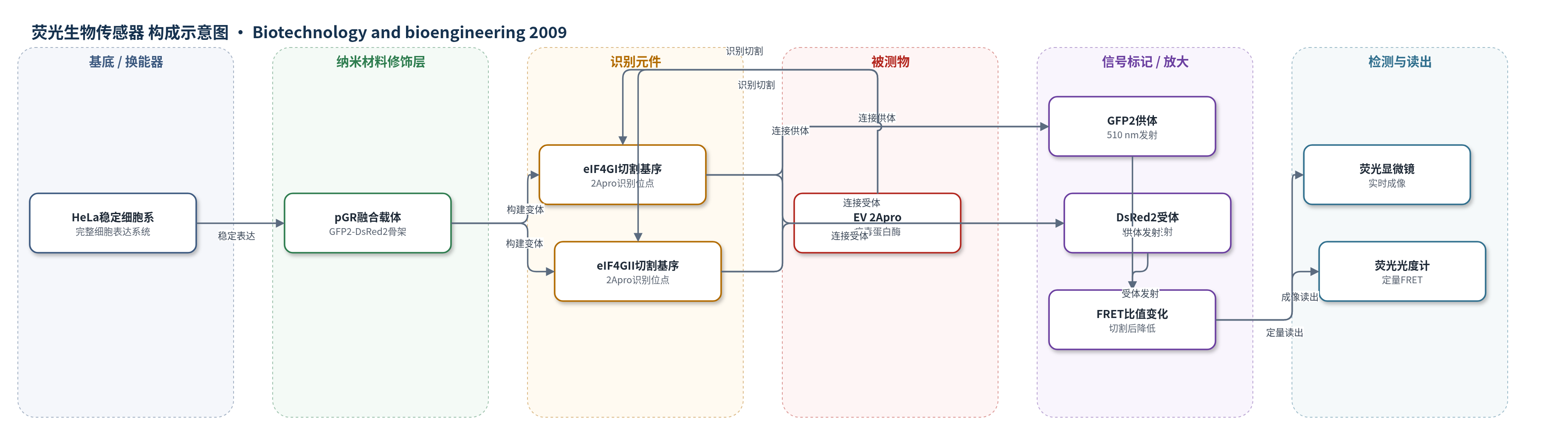
点击图片查看大图 · 依据论文自动绘制
传感器类型
荧光生物传感器
检测对象
肠道病毒 2A 蛋白酶(enterovirus 2A protease, 2Apro)对 eIF4GI/eIF4GII 的切割活性;样品基质:HeLa 活细胞(EV71、CVA16、CVB1、CVB4 感染细胞)
检测原理
该传感器由 GFP2 供体、eIF4G 切割基序和 DsRed2 受体串联组成。未切割时,GFP2 与 DsRed2 空间邻近,390 nm 激发后供体能量以非辐射方式转移至受体,产生 590 nm 受体发射,FRET 比值(590/510 nm)较高。当肠道病毒 2Apro 进入细胞并识别 eIF4GI 或 eIF4GII 切割基序时,在特定位点水解融合蛋白,使供体与受体分离,FRET 效率下降,表现为受体发射降低、供体发射升高,FRET 比值随 2Apro 活性/病毒滴度增加而下降。DsRed2 寡聚化使完整融合蛋白滞留胞质,切割后单聚 GFP2 可进入细胞核,提供空间分辨标记。系统通过荧光显微镜成像或荧光光度计定量 FRET 比值,从而实时反映 2Apro 对 eIF4G 的切割动力学。
检测灵敏度
原文未报告 LOD、线性范围、灵敏度斜率或 R^2;检测范围: 0.04–5 PFU/cell;最低可分辨滴度: 0.04 PFU/cell at 36 h or 0.2 PFU/cell at 18 h p.i.
效应效果
该 FRET 生物传感器特异性高:FRET 降低仅由 2Apro 催化活性引起,单独表达 3Cpro、失活 2Apro 或空载体均不破坏 FRET;MPCMK 可剂量依赖逆转 FRET 降低,Z-VAD-FMK、cisplatin 和 cyclohexamide 不改变 FRET 信号。FRET 比值与融合底物切割比例及内源 eIF4GI/eIF4GII 切割比例高度相关(P < 0.001),可区分 eIF4GI 早于 eIF4GII 的切割动力学。检测范围为 0.04–5 PFU/cell,0.04 PFU/cell 感染 36 h 或 0.2 PFU/cell 感染 18 h 即可分辨。传感器适用于 EV71、CVA16、CVB1 和 CVB4,HSV-1 感染不引起 FRET 下降,作者认为其可作为研究病毒诱导宿主翻译抑制和筛选抗肠道病毒化合物的通用平台。
传感器的构成
- 表达载体/换能骨架:pGR 质粒,编码 GFP2-DsRed2 融合蛋白与 zeocin 抗性,用于在 HeLa 细胞中稳定表达 FRET 对。
- 供体荧光蛋白:GFP2,作为 FRET 供体,390 nm 激发并在 510/515 nm 发射。
- 受体荧光蛋白:DsRed2,作为 FRET 受体,540 nm 激发并在 590/600 nm 发射,与 GFP2 形成 FRET 对。
- 识别/切割元件:eIF4GI 切割基序(residues 675–686)或 eIF4GII 切割基序(residues 693–704),连接 GFP2 与 DsRed2,被 2Apro 特异性切割。
- 突变对照元件:eIF4GImut/eIF4GIImut 切割基序,P10 位点 G→R 突变,用于验证 2Apro 切割特异性。
- 细胞表达基质:HeLa 稳定细胞系 FRETwt1/FRETmut1/FRETwt2/FRETmut2,提供完整细胞内环境与内源 eIF4G 背景。
- 信号读出装置:Nikon TE200 倒置荧光显微镜与 Fluoroskan Ascent 374 荧光光度计,390 nm 激发、510/590 nm 发射,计算 FRET 比值。
中文摘要
肠道病毒感染可显著关闭宿主蛋白合成,该过程主要由病毒 2A 蛋白酶(2Apro)切割真核翻译起始因子 eIF4GI 和 eIF4GII 介导。本研究基于荧光共振能量转移(FRET)开发了遗传编码的 FRET 生物传感器,用于在完整细胞中可视化和定量该特异性蛋白水解过程。通过稳定表达由绿色荧光蛋白 2(GFP2)和红色荧光蛋白 2(DsRed2)组成的融合底物,并在两者之间连接 eIF4GI 或 eIF4GII 的 2Apro 切割基序,实现了 FRET 信号对切割事件的响应。感染肠道病毒后,FRET 生物传感器显示实时且可定量的 FRET 降低;FRET 降低水平与内源 eIF4G 异构体的切割动力学密切相关。FRET 损伤仅归因于 2Apro 催化活性,而不受其他病毒编码蛋白酶、激活的 caspase 或感染细胞中蛋白合成普遍抑制的影响。该 FRET 生物传感器可作为多种相关肠道病毒的通用平台。FRET 提供的时空与定量成像有助于在正常细胞环境中研究蛋白酶–底物行为,从而阐明病毒诱导宿主翻译抑制的分子机制。
英文摘要
Enterovirus (EV) infection has been shown to cause a marked shutoff of host protein synthesis, an event mainly achieved through the cleavages of eukaryotic translation initiation factors eIF4GI and eIF4GII that are mediated by viral 2A protease (2A(pro)). Using fluorescence resonance energy transfer (FRET), we developed genetically encoded and FRET-based biosensors to visualize and quantify the specific proteolytic process in intact cells. This was accomplished by stable expression of a fusion substrate construct composed of the green fluorescent protein 2 (GFP(2)) and red fluorescent protein 2 (DsRed2), with a cleavage motif on eIF4GI or eIF4GII connected in between. The FRET biosensor showed a real-time and quantifiable impairment of FRET upon EV infection. Levels of the reduced FRET closely correlated with the cleavage kinetics of the endogenous eIF4Gs isoforms. The FRET impairments were solely attributed to 2A(pro) catalytic activity, irrespective of other viral-encoded protease, the activated caspases or general inhibition of protein synthesis in the EV-infected cells. The FRET biosensors appeared to be a universal platform for several related EVs. The spatiotemporal and quantitative imaging enabled by FRET can shed light on the protease-substrate behaviors in their normal milieu, permitting investigation into the molecular mechanism underlying virus-induced host translation inhibition.