组成图示
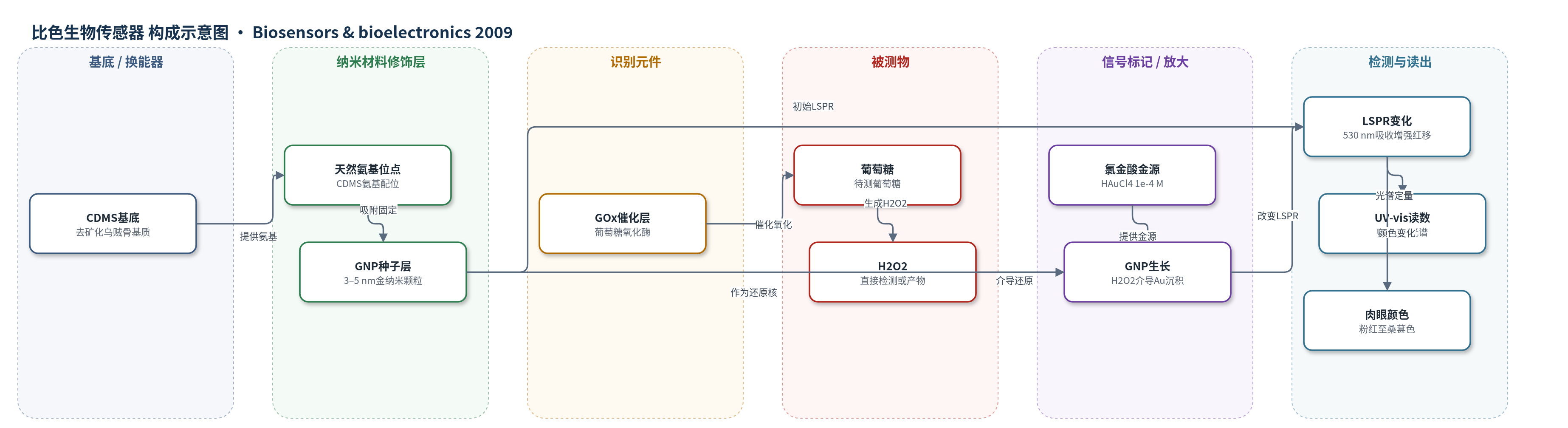
点击图片查看大图 · 依据论文自动绘制
传感器类型
比色生物传感器
检测对象
过氧化氢(hydrogen peroxide, H2O2)、葡萄糖(glucose);样品基质为磷酸盐缓冲液(PBS,pH 7.4,0.02 M),葡萄糖检测体系含GOx和HAuCl4。
检测原理
该传感器以CDMS负载的GNPs作为光学换能元件。对于H2O2直接检测,预吸附的GNPs作为无电沉积的成核/催化位点,H2O2将溶液中的HAuCl4还原为Au0并沉积在GNPs表面,使GNPs尺寸增大;GNPs尺寸增大导致其LSPR吸收峰强度增强并发生红移,UV–vis在530 nm处的吸光度随H2O2浓度升高而增大。对于葡萄糖检测,GOx催化葡萄糖氧化生成H2O2,随后触发相同的HAuCl4还原和GNPs生长过程,因此吸光度变化间接反映葡萄糖浓度。CDMS高比表面积可固定大量GNPs,通过纳米颗粒尺寸变化放大光学响应。
检测灵敏度
H2O2: LOD: 2 × 10⁻⁶ M;检测范围: 2 × 10⁻⁶–1.5 × 10⁻⁴ M。Glucose: 线性范围: 5 × 10⁻⁶–5 × 10⁻⁵ M;线性方程: Abs = 0.30517 + 0.08238 c (10−5 M);R = 0.99807;n = 6。
效应效果
作者设置无H2O2或葡萄糖对照,但未报告选择性/抗干扰数据;三次重复给出误差棒,未报告RSD。CDMS 4 ℃可保存至少2个月,金功能化CDMS 4 ℃蒸馏水中稳定数天。与玻璃基底相比,本文响应更高:H2O2 2×10⁻⁵–7.2×10⁻³ M时吸光度约0.3–1.0,玻璃约0.16–0.26;葡萄糖5×10⁻⁶–3×10⁻⁴ M时本文约0.30–0.73,玻璃约0.17–0.29。H2O2检出限2×10⁻⁶ M低于玻璃的2×10⁻⁵ M。TEM显示5×10⁻⁵ M葡萄糖反应20 min后GNPs增至约12 nm。作者认为其简单快速,可肉眼判色,适合定性或半定量初筛。
传感器的构成
- 基底/换能器:乌贼骨来源基质(cuttlebone-derived matrix substrate, CDMS),去矿化透明有机/几丁质基质,三维腔室结构、高比表面积,直接承载GNPs并作为光学透明基底
- 氨基结合层:CDMS天然氨基(-NH2)位点,通过强配位作用直接吸附固定GNPs,无需额外化学修饰
- 纳米材料修饰层:金纳米颗粒(gold nanoparticles, GNPs,3–5 nm金种子),作为LSPR光学换能元件和后续金沉积的催化核
- 识别/催化元件:葡萄糖氧化酶(glucose oxidase, GOx,12 μg/mL),用于葡萄糖检测,催化葡萄糖氧化生成H2O2
- 信号放大试剂:氯金酸(HAuCl4,1×10−4 M)与H2O2/GOx体系,H2O2介导HAuCl4还原为Au0并沉积在GNPs表面,使GNPs增大
- 缓冲介质:磷酸盐缓冲液(PBS,pH 7.4,0.02 M),维持反应环境
- 信号读出:UV–vis吸收光谱(Shimadzu UV3150,400–850 nm,530 nm处定量)及肉眼颜色变化(粉红至桑葚色)
中文摘要
本文报道了一种基于乌贼骨来源基质(CDMS)和金纳米颗粒(GNPs)的简单光学生物传感器,用于过氧化氢(H2O2)和葡萄糖检测。CDMS具有三维腔室结构,天然富含氨基,可在无需复杂修饰的情况下直接固定GNPs;其高比表面积可负载大量GNPs,从而增强光学信号转换并提高检测灵敏度。传感器原理是:固定于CDMS上的GNPs的局域表面等离子共振(LSPR)吸收会随H2O2介导氯金酸化学还原、使GNPs尺寸增大而发生显著变化。作者以UV–vis吸收光谱获取H2O2或葡萄糖浓度的定量信息。H2O2检测范围为2×10⁻⁶至1.5×10⁻⁴ M,葡萄糖线性响应范围为5×10⁻⁶至5×10⁻⁵ M。此外,GNPs在CDMS上的生长可引起肉眼可见的颜色变化,使该传感器在医学和生物技术领域的定性或半定量快速筛查中具有潜在应用价值。
英文摘要
A simple biosensor for hydrogen peroxide (H(2)O(2)) and glucose was fabricated by incorporating gold nanoparticles (GNPs) onto a cuttlebone-derived matrix substrate (CDMS). Such a three-dimensional chamber-like structure naturally bears abundant amino groups for the direct immobilization of GNPs without a series of modifications. And preferably, the framework endows CDMS with a very high surface area for the attachment of GNPs, resulting in effective optical signal transduction and improved sensitivity of the detection system. The principle behind this biosensor is that the localized surface plasmon resonance (LSPR) of the immobilized GNPs changes with the enlargement of GNPs by H(2)O(2)-mediated chemical reduction of chloroauric acid. Using this approach, we demonstrate the proof of an optical biosensor to quantify the concentration of H(2)O(2) as well as glucose. UV-vis absorption spectra were recorded to obtain quantitative information about the H(2)O(2) or glucose concentration. The detection range of our biosensor to H(2)O(2) concentration was from 2 x 10(-6) to 1.5 x 10(-4)M, while the linear response range of glucose concentration was from 5 x 10(-6) to 5 x 10(-5)M. Inspiringly and interestingly, the growth of GNPs on CDMS gives rise to color changes, this phenomenon shows that the rapid detection by our sensor has the superiority in visual detection to a certian extent, which has been a potential application in qualitative or semiquantitative analysis for medicine and biotechnology.