组成图示
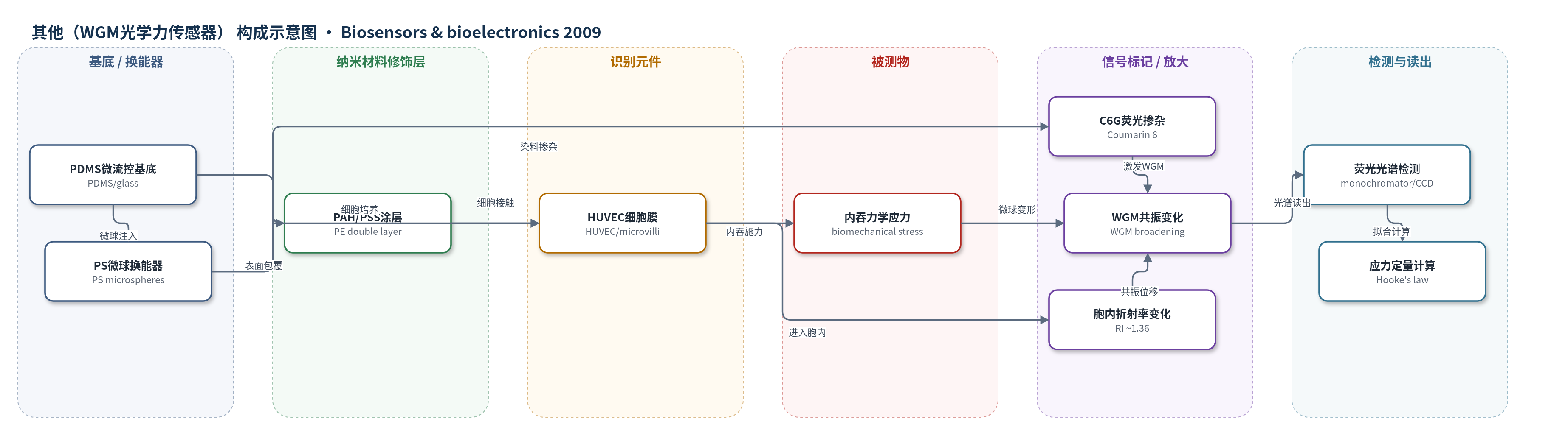
点击图片查看大图 · 依据论文自动绘制
传感器类型
其他(WGM光学力传感器)
检测对象
细胞内吞生物力学应力(biomechanical stress/cortical tension,含主动应力分量);样品基质:人脐静脉内皮细胞(HUVECs)/内皮细胞生长培养基(ECGM)微流控体系
检测原理
荧光掺杂 PS 微球本身构成 WGM 光学谐振腔。HeCd 激光激发 C6G 染料后,其各向荧光发射可同时激发三维 WGM。当 HUVEC 接触并内吞微球时,细胞膜与细胞骨架对微球施加面内和面外应力,使球形微球弹性变形为椭球;同时微球进入胞内,环境折射率升高。变形解除 WGM 极化简并,引起共振展宽和蓝移,折射率变化引起共振位移。单色仪与 CCD 记录荧光 WGM 光谱,经 Lorentzian 拟合和 Airy 近似提取平均折射率及最小/最大半径,再结合涂层厚度和广义 Hooke 定律计算应力。应力越大,共振展宽与位移越显著。
检测灵敏度
原文未报告 LOD、线性范围、灵敏度斜率或相关系数。
效应效果
该方法可在活细胞内吞全程实时、非破坏地监测微球变形,WGM 峰拟合偏差通常小于0.006–0.03 nm,整体拟合偏差约0.1 nm以内。对7.8 μm微球,5 min时面内应力为-1.282±0.593 MPa、面外应力为0.843±0.279 MPa,对应面外总力约40.2 μN,显著高于被动皮层张力(约200 kPa)和表面分子马达估计(约95 nN),表明存在细胞骨架主动应力;10.1 μm微球应力比7.8 μm微球小5–6倍。作者认为误差主要来自涂层厚度和材料常数,而非换能机制,方法可推广至其他吞噬细胞、炎症免疫和癌症力学表型研究。
传感器的构成
- 微球换能器:聚苯乙烯微球(PS microspheres,6–10 μm),作为 WGM 谐振腔与力学探针,被细胞内吞并发生弹性变形。
- 荧光激发/信号标记:香豆素6(Coumarin 6, C6G)染料掺杂于 PS 微球内部,受激光激发后向各向发射,激发三维 WGM 并提供荧光光谱读出。
- 表面修饰层:聚电解质双层(PAH/PSS,poly(allylamine hydrochloride)/poly(sodium 4-styrene-sulfonate)),层层自组装包覆微球,形成薄表面层并影响细胞接触。
- 细胞识别/力学作用层:人脐静脉内皮细胞(HUVECs)细胞膜与微绒毛,接触并内吞微球,施加皮层与细胞骨架应力。
- 微流控基底:PDMS(Sylgard 184)微流控流道与玻璃盖玻片(涂 PAH/PSS 促进 HUVEC 黏附),承载 ECGM 和 HUVECs,提供静止观察环境。
- 光学读出系统:HeCd 激光(441.6 nm)、倒置显微镜、单色仪与 CCD,激发微球并记录 WGM 荧光光谱。
中文摘要
本文报道了活细胞内吞过程中生物力学应力的直接测量方法。作者将荧光染料掺杂的聚苯乙烯微球作为微型远程光学传感器,利用回音壁模式(WGM)作为换能机制。在微球被人脐静脉内皮细胞逐步内吞的过程中,通过连续监测 WGM 共振,可以确定微球所经历的机械变形,其表现为共振峰展宽和蓝移,并据此推算细胞对微球施加的应力。结果表明,内吞过程中出现的应力出乎意料地高,最大可达被动皮层张力的五倍;该现象无法仅用被动皮层张力解释,而只能归因于细胞骨架机器在颗粒内吞过程中产生的主动应力分量。该方法可推广至其他吞噬细胞,为细胞生物学、炎症免疫学和癌症研究提供了一种新型生物力学研究工具。
英文摘要
Direct measurement of the biomechanical stress induced by a live cell during endocytosis is reported. Fluorescent dye-doped polystyrene microspheres were used as microscopic remote optical sensors applying whispering gallery modes (WGMs) as transducer mechanism. Monitoring of the WGMs throughout the incorporation of the microsphere into the cell enabled the determination of the deformation experienced by the microsphere, characterized by both a broadening and a blue shift of the resonances, and consequently the stress induced by the cell. The results reveal an unexpectedly high stress with a magnitude of up to five times that of the passive cortical tension, which can be only explained by a so far undetermined active stress component induced by the cytoskeletal machinery during particle incorporation. The method is adaptable to the study of any other kind of phagocyte and thus provides a novel research tool of high interest for cell biology.