组成图示
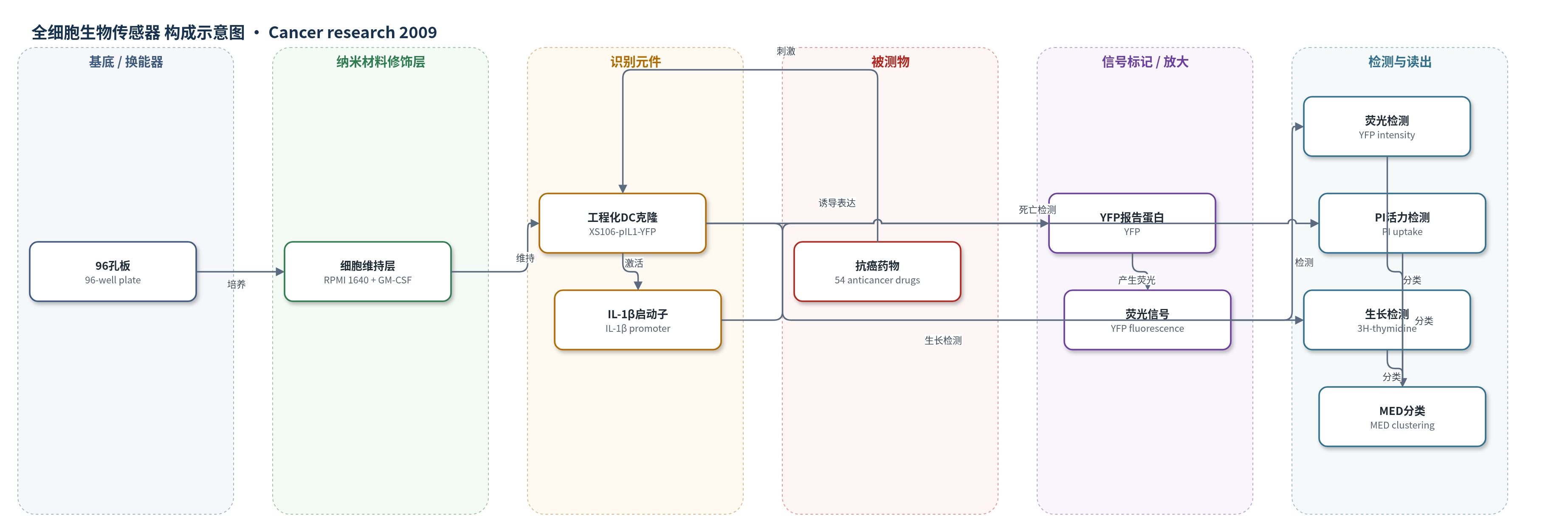
点击图片查看大图 · 依据论文自动绘制
传感器类型
全细胞生物传感器
检测对象
抗癌药物(chemotherapeutic agents / anticancer drugs),样品基质为细胞培养液(RPMI 1640 + GM-CSF + NS conditioned medium,DMSO 0.5%)
检测原理
该全细胞生物传感器以工程化DC克隆XS106-pIL1-YFP为识别与响应单元。抗癌药物加入细胞培养体系后,与DC表面或胞内靶点相互作用,触发DC成熟相关信号通路,使IL-1β启动子激活并驱动YFP转录表达。YFP荧光强度随药物浓度呈剂量依赖性升高,反映药物诱导DC成熟的相对效力。系统同时以PI摄取评估细胞死亡、以3H-胸苷掺入评估GM-CSF依赖生长,从而获得成熟、存活和生长三项剂量-反应曲线。通过计算最小有效剂量(MED)并进行聚类,可将药物区分为DC刺激性、生长抑制性、毒性或无显著作用类型。
检测灵敏度
未报告 LOD、线性范围、灵敏度斜率或 R^2。
效应效果
研究对54种抗癌药物在5个浓度、每孔复孔条件下筛选,实验至少重复三次,未报告RSD、回收率或与传统方法对比。基于MED聚类将药物分为4类:15种为type 1(亚细胞毒性诱导DC成熟),19种主要抑制生长,1种引起生长停滞和死亡而无成熟信号,19种无显著变化。长春碱表现最突出,可上调CD40、CD80、CD86和MHC II,诱导IL-1β、IL-6和IL-12,增强同种异体T细胞刺激能力,并使FITC-葡聚糖摄取提高约10倍,增强OVA蛋白向OT-I CD8 T细胞的交叉呈递。作者认为该结果可为化疗药物的免疫学选择和联合应用提供依据。
传感器的构成
- 反应容器/培养基底:96孔板(96-well plate),承载细胞与药物孵育。
- 细胞维持层:完全RPMI 1640培养基,含0.5 ng/mL GM-CSF和5% NS细胞条件培养基,维持XS106 DC存活与生长。
- 全细胞识别/响应元件:工程化小鼠树突状细胞克隆XS106-pIL1-YFP DC clone,作为药物刺激的生物传感器细胞。
- 报告基因调控元件:IL-1β启动子(IL-1β promoter),响应DC成熟信号并驱动下游报告基因。
- 荧光信号元件:黄色荧光蛋白(YFP),表达量反映IL-1β启动子激活程度。
- 辅助活力读出:碘化丙啶(PI),标记膜完整性受损的死亡细胞,用于评估药物细胞毒性。
- 辅助生长读出:3H-胸苷(3H-thymidine),掺入DNA以检测GM-CSF依赖生长。
中文摘要
树突状细胞(DC)在抗肿瘤免疫中起关键作用,但许多化疗药物对其药理学效应仍不清楚。作者此前通过工程化小鼠DC系XS106,使其在白细胞介素-1β(IL-1β)启动子控制下稳定表达黄色荧光蛋白(YFP),构建了DC生物传感器克隆。本研究利用XS106-pIL1-YFP DC克隆对54种抗癌药物进行无偏筛选,每种药物在0.1–10 μmol/L五个浓度下检测YFP表达、细胞活力和粒细胞-巨噬细胞集落刺激因子(GM-CSF)依赖生长。系统筛选显示药物对三项功能变量存在显著异质性。15种药物在亚细胞毒性浓度诱导显著YFP表达,被归为“DC刺激性”抗癌药物,并进一步证实可诱导小鼠骨髓来源DC至少一种特征性成熟改变。例如长春碱诱导IL-1β、IL-6和IL-12产生,上调CD40、CD80、CD86和MHC II表面表达,并增强DC刺激T细胞的能力。这些结果揭示了常用化疗药物对DC的差异化药理效应,可为临床合理选择和联合抗癌药物提供概念框架。
英文摘要
Despite the crucial roles dendritic cells (DC) play in host immunity against cancer, the pharmacologic effects of many chemotherapeutic agents have remained mostly unknown. We recently developed a DC biosensor clone by engineering the stable murine DC line XS106 to express the yellow fluorescent protein (YFP) gene under the control of interleukin (IL)-1beta promoter. In this study, the resulting XS106 pIL1-YFP DC clone was used to screen 54 anticancer drugs. Each drug was tested at five concentrations (0.1-10 micromol/L) for its effects on YFP expression, cell viability, and granulocyte macrophage colony-stimulating factor-dependent growth. Our unbiased systematic screening unveiled a striking heterogeneity among the tested anticancer drugs in their effects on the three functional variables. Interestingly, 15 drugs induced significant YFP expression at subcytotoxic concentrations and were thus categorized as "DC-stimulatory" anticancer drugs. These drugs were subsequently found to induce at least one of the characteristic maturational changes in mouse bone marrow-derived DCs. For example, vinblastine, a prototypic drug of this class, induced the production of IL-1beta, IL-6, and IL-12, increased surface expression of CD40, CD80, CD86, and MHC class II, and augmented T cell-stimulatory capacity of DCs. Not only do these results illustrate the differential pharmacologic effects of commonly used chemotherapeutic agents on DCs, they may also provide a conceptual framework for rationale-based selection and combination of anticancer drugs for clinical application.