组成图示
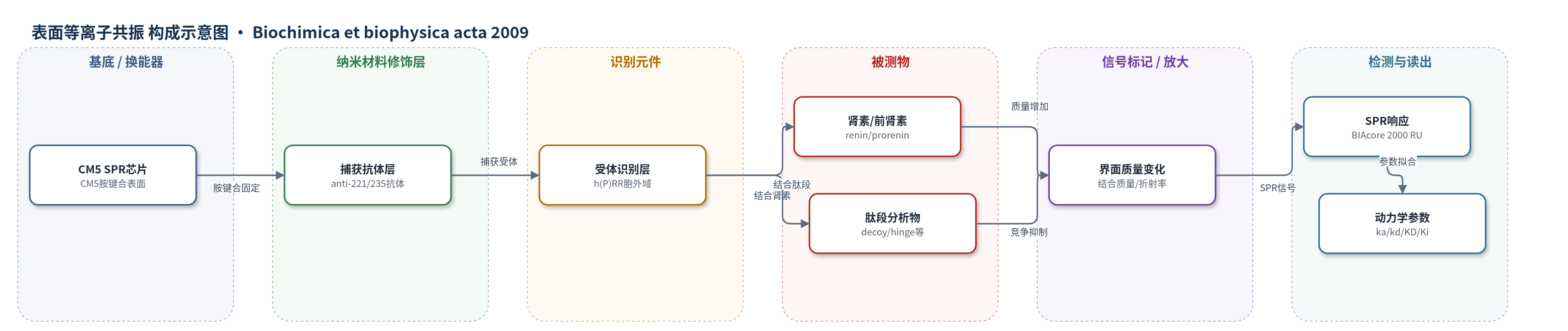
点击图片查看大图 · 依据论文自动绘制
传感器类型
表面等离子共振(SPR)生物传感器
检测对象
人肾素(human renin)、人前肾素(human prorenin)、decoy肽(R10PIFLKRMPSI19P)、hinge肽(S149QGVLKEDVF158)、1P-8P(L1PPTDTTTF8P)、1P-15P(L1PPTDTTTFKRIFLKR15P);样品基质为HBS-EP缓冲液(SPR)或PBS(塑料孔平衡)
检测原理
该检测基于表面等离子共振(SPR)换能。anti-(P)RR抗体anti-221/235经胺键合固定在CM5芯片上,再捕获重组人(P)RR胞外域,形成受体识别界面。肾素、前肾素或肽段流经芯片时与h(P)RR结合,使界面质量/折射率增加,SPR共振角偏移,BIAcore 2000以resonance units(RU)实时记录结合与解离曲线。结合量随分析物浓度升高而增加并趋于饱和;用Langmuir 1:1模型拟合ka、kd和KD。decoy或hinge肽与受体竞争结合后降低肾素/前肾素的RU响应,按KDapp=KD×(1+[i]/Ki)计算Ki。
检测灵敏度
—
效应效果
抗体介导固定使h(P)RR保持可结合构象,肾素、前肾素及肽段产生可重复SPR响应。平衡法KD:肾素4.5±1.5 nM、前肾素1.0±0.1 nM;BIAcore法:4.4±1.8和1.2±0.4 nM,前肾素亲和力约为肾素4倍。decoy肽KD 3.5±1.6 nM,hinge肽17.0±2.6 nM。decoy与hinge肽对肾素/前肾素结合的Ki分别为16.7±3.5/15.1±2.8和37.1±6.2/30.7±3.1 nM。再生缓冲液可恢复表面结合能力,Western blot证实前肾素结合后完整且decoy降低结合。80 nM肽段不抑制肾素酶活,Km约0.1 μM。作者认为该SPR系统可用于(P)RR研究,支持前肾素至少两个高亲和位点、肾素一个位点。
传感器的构成
- 基底/换能器:CM5 SPR芯片(胺键合表面),提供表面等离子共振换能界面
- 捕获抗体层:anti-(P)RR抗体anti-221/235(兔多克隆抗体,识别E221IGKRYGEDSEQFRD235表位),胺键合固定并捕获受体
- 识别元件层:重组人(P)RR胞外域h(P)RR(N17-N308,6×His tag,32.5 kDa),与肾素/前肾素/肽段特异性结合
- 分析物:human renin、human prorenin、decoy肽R10PIFLKRMPSI19P、hinge肽S149QGVLKEDVF158、1P-8P、1P-15P等,结合后改变界面质量
- 运行缓冲液:HBS-EP(0.15 M NaCl、3.0 mM EDTA、0.005% polysorbate 20、0.01 M HEPES,pH 7.4),维持结合环境
- 再生缓冲液:10 mM glycine与150 mM NaCl,洗脱配体并再生抗体表面
- 信号读出:BIAcore 2000 SPR仪器,以resonance units(RU)实时监测结合质量变化
中文摘要
近期研究提示decoy肽序列参与前肾素与(pro)肾素受体[(P)RR]的结合。本研究利用“hinge”肽S149QGVLKEDVF158、L1PPTDTTTF8P、L1PPTDTTTFKRIFLKR15P及decoy肽R10PIFLKRMPSI19P,探讨肾素和前肾素中与(P)RR相互作用的关键区域。动力学分析中,重组人(P)RR经特异性抗(P)RR抗体固定在生物传感器表面;平衡分析中,(P)RR直接吸附于塑料孔板。肾素和前肾素与预吸附受体的KD分别为4.5和1.0 nM,与BIAcore结果相近。“hinge”肽剂量依赖结合(P)RR,KD约17.0 nM,约为decoy的5倍;L1PPTDTTTF8P和L1PPTDTTTFKRIFLKR15P的KD分别为52和7.6 nM。“hinge”肽与decoy肽均抑制肾素和前肾素结合,Ki分别为16.7和15.1 nM、37.1和30.7 nM。结果提示肾素有一个、前肾素至少有两个高亲和结合位点。
英文摘要
An important role of the decoy peptide sequence has recently been suggested in vitro for the binding of prorenin to the (pro)renin receptor [PRR]. In this study, other prospective crucial regions in renin and prorenin responsible for their interaction with PRR were investigated using various kinds of peptides, e.g., the "hinge" S149QGVLKEDVF158 designed from the structure of renin also common to prorenin, L1PPTDTTTF8P, L1PPTDTTTFKRIFLKR15P and the decoy (R10PIFLKRMPSI19P) designed from the predicted structure of prorenin. For the kinetic analysis, the recombinant hPRR was immobilized on the biosensor surface through a specific anti-PRR antibody. In case of the equilibrium state analysis, the PRR was directly adsorbed on plastic wells for observing the bindings of renin/prorenin. The dissociation constants (KD) for the bindings of renin and prorenin to the pre-adsorbed receptors were 4.5 and 1.0 nM, respectively, similar to those stated in the kinetic study by BIAcore assay. The "hinge" region peptide bound to PRR in a dose-dependent manner with a KD estimated 17.0 nM which was five times higher than that of the decoy. The KD values for L1PPTDTTTF8P and L1PPTDTTTFKRIFLKR15P were 52 and 7.6 nM, respectively. The "hinge" peptide, as the decoy, inhibited the bindings of renin and prorenin to PRR. The inhibition constant (Ki) for the binding of renin and prorenin by the decoy and "hinge" were 16.7 and 15.1, and 37.1 and 30.7 nM, respectively. These in vitro studies suggest that renin has a single and prorenin has at least two high affinity binding sites for the PRR.