组成图示
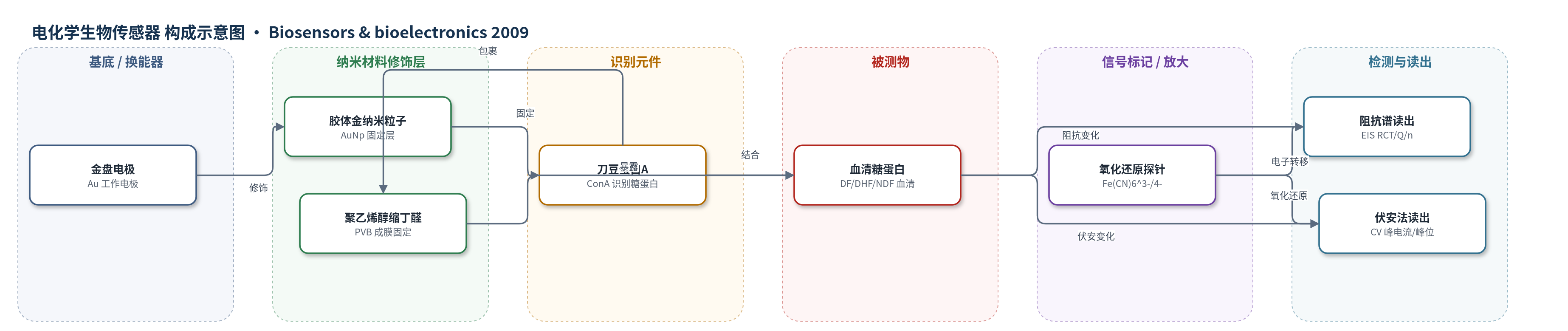
点击图片查看大图 · 依据论文自动绘制
传感器类型
电化学生物传感器
检测对象
登革热(dengue fever, DF)血清糖蛋白(serum glycoproteins)、登革出血热(dengue hemorrhagic fever, DHF)血清糖蛋白、阴性(non-dengue fever, NDF)血清糖蛋白;样品基质:人血清(PBS 1:30 稀释)
检测原理
传感器以金电极为换能器,AuNp 提供高比表面积并固定 ConA,PVB 将 AuNp–ConA 聚集体包裹固定,BSA 封闭非特异性位点。检测时,DF/DHF/NDF 血清中的糖蛋白与 ConA 的糖基识别位点结合,形成 ConA–糖蛋白复合物并在电极界面聚集。该复合物形成电子转移阻挡层,阻碍 K3[Fe(CN)6]/K4[Fe(CN)6] 探针在电极表面的电子转移,使 CV 峰电流下降、峰位分离增大,响应由扩散控制转向不可逆/电子转移控制;EIS 中电荷转移电阻 RCT 增大,Q 和 n 随界面电容与分散特性变化。DF、DHF 和 NDF 血清因糖蛋白模式不同,产生不同 RCT/Q/n 组合,可用相对 RCT 变化或三维图区分。
检测灵敏度
未报告 LOD、线性范围、灵敏度斜率、相关系数。
效应效果
传感器对 DF、DHF、NDF 血清呈现可区分阻抗响应。ΔRCT 分别为 DF 162.2 ± 21.3%、DHF 136.3 ± 30.9%、NDF 41.1 ± 4.6%;DF 与 DHF 在 90% 置信水平无统计差异,但均高于 NDF。三维 RCT–Q–n 图清晰分离三类血清:NDF 位于低 RCT/Q、高 n 区,DF 位于高 RCT、低 Q/n 区。TEM 显示 DHF 血清引起 AuNp 聚集,支持 ConA 结合糖蛋白。每类血清 3 例患者、每例 3 次重复,显示一定重现性;未报告长期稳定性、抗干扰、加标回收率或与 ELISA/PCR 对比。作者认为该电极及三维阻抗分析可用于识别血清糖蛋白模式,辅助登革热免疫反应诊断。
传感器的构成
- 工作电极/基底:金盘电极(gold disc electrode, d=2 mm),机械抛光后作为电化学换能器。
- 纳米材料修饰层:胶体金纳米粒子(AuNp,HAuCl4/柠檬酸钠还原制备,UV-vis 527 nm),提供高比表面积并固定 ConA。
- 识别元件:刀豆蛋白A(Concanavalin A, ConA, 200 μg/mL),识别血清糖蛋白糖基化位点。
- 固定/成膜层:聚乙烯醇缩丁醛(PVB, 2% v/v 乙醇溶液),包裹 AuNp–ConA 聚集体并固定于电极表面。
- 封闭剂:牛血清白蛋白(BSA, 0.2% PBS, 37 °C 20 min),封闭剩余活性位点。
- 氧化还原探针:K3[Fe(CN)6]/K4[Fe(CN)6](1:1, 10 mM),用于 CV/EIS 电子转移信号读出。
中文摘要
本研究将刀豆蛋白A(Concanavalin A, ConA)通过金纳米粒子(AuNp)和聚乙烯醇缩丁醛(PVB)固定于金电极表面,并利用循环伏安法(CV)和电化学阻抗谱(EIS)进行表征。以含10 mM K3[Fe(CN)6]/K4[Fe(CN)6](1:1)的磷酸盐缓冲液作为氧化还原探针,在100 mHz–100 kHz频率范围内进行EIS,在−0.2–0.7 V(vs. Ag/AgCl)范围内进行CV。将传感器分别暴露于登革热(DF)、登革出血热(DHF)及阴性(NDF)患者血清后,伏安响应由明显扩散控制逐渐变为不可逆行为;EIS显示感染血清使电子转移电阻显著升高。通过等效电路拟合获得电荷转移电阻和电容参数,并利用相对电荷转移电阻变化及三维阻抗图区分DF、DHF和NDF血清。不同阻抗模式归因于血清中糖蛋白模式差异。结果表明,AuNp–ConA–PVB修饰电极结合三维阻抗分析可作为识别疾病免疫反应中糖蛋白模式的潜在电化学生物传感器。
英文摘要
Immobilization of concanavalin A on gold electrode by means of gold nanoparticles and polyvinyl butyral was carried out and investigated by cyclic voltammetry and electrochemical impedance spectroscopy. The system was tested with sera from patients infected by dengue fever (DF) and dengue hemorrhagic fever (DHF). Electrochemical impedance spectroscopy (in the frequency range from 100mHz to 100KHz), and cyclic voltammetry (from -0.2 to 0.7V vs. Ag/AgCl), was performed in phosphate buffer solution containing 10mM K(3)[Fe(CN)(6)]/K(4)[Fe(CN)(6)] (1:1) mixture as a redox probe. As biomolecules accumulated on the electrode surface the voltammetric response changed from a clear diffusional to an irreversible behavior. Impedance spectroscopy showed a clear increase of the electron-transfer resistance when the sensor is exposed to contaminated sera (DF or DHF) as compared to exposure to uncontaminated serum (NDF). The results were analyzed through an equivalent circuit and values of charge transfer resistance and capacitance were obtained. Variations in charge transfer resistance were used to distinguish the sensor response for the different sera investigated (DF, DHF and NDF). Alternatively, a three-dimensional graph gave the best response for differentiation of all three blood sera. The distinctive patterns of impedimetric responses observed were ascribed to different glycoprotein patterns in the sera investigated. Therefore, the lectin immobilization on electrode surface with gold nanoparticles and polyvinyl butyral, combined with the three-dimensional impedance analysis introduced herein are valuable tools in the development of a biosensor for immunological response to diseases.