组成图示
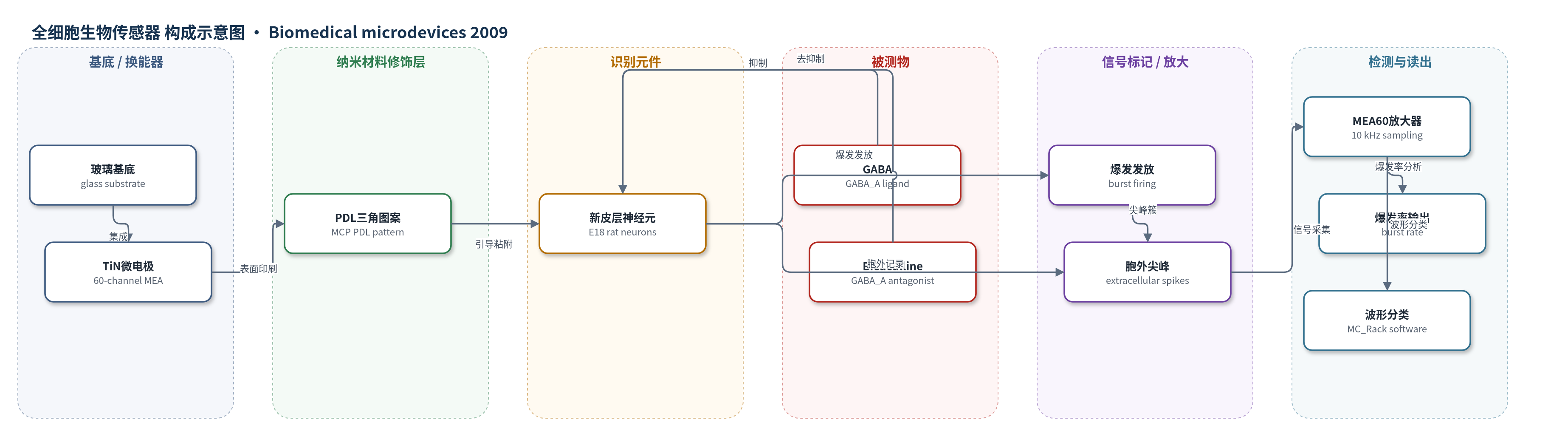
点击图片查看大图 · 依据论文自动绘制
传感器类型
全细胞生物传感器
检测对象
γ-氨基丁酸(GABA)、比库替啶(Bicuculline);样品基质:神经元培养液(B27/Neurobasal培养基)
检测原理
微接触印刷在TiN微电极表面形成三角形PDL粘附图案,使神经元细胞体优先粘附于电极节点,突起沿图案线条延伸并分支,增强细胞-电极耦合并构建可重复的低密度网络。神经元作为细胞基传感元件,其突触网络产生自发胞外尖峰和同步爆发。当GABA结合突触后GABA_A受体时,增强氯离子内流和抑制性突触后电流,降低神经元兴奋性,使爆发率随GABA浓度增加而下降;Bicuculline阻断GABA_A受体,解除GABA能抑制,使爆发率随浓度增加而上升。MEA多通道采集胞外电位,MC_Rack软件检测尖峰并统计爆发率,从而将分析物浓度转换为电生理剂量-反应信号。
检测灵敏度
IC50(GABA): 1.63±0.14 µM;IC50(GABA, random): 1.43±0.11 µM;EC50(Bicuculline): 0.77±0.11 µM;EC50(Bicuculline, random): 0.80±0.14 µM
效应效果
图案化网络在200 cells/mm²低密度下平均覆盖28个电极,随机网络仅11个,电极覆盖提高约三倍;电活动可从约25/60个电极记录,随机低密度仅10个。尖峰信噪比大于5:1,负成分通常50–100 µV,峰-峰通常大于100 µV。成熟网络出现稳定同步爆发,频率3–6次/分钟,持续100–150 ms,爆发内尖峰频率约150 Hz,静默期至少0.5 s,并维持至少4周。GABA呈剂量依赖性抑制爆发,10 µM时完全抑制;Bicuculline引起去抑制,撤除后10 min内恢复。形态和突触密度与随机网络相似,作者认为其适合用作细胞基生物传感器。
传感器的构成
- 基底/换能器电极:玻璃基底上的60通道钛氮化钛(TiN)微电极阵列(MEA),电极直径30 µm、间距200 µm,用于胞外电记录
- 表面图案化粘附层:微接触印刷(MCP)沉积的三角形多聚-D-赖氨酸(PDL)图案,线宽6 µm,节点与电极坐标匹配,引导细胞体粘附于电极并引导突起沿线条生长
- 识别/传感元件:原代大鼠新皮层神经元(neocortical neurons,E18大鼠胎儿皮层细胞),作为细胞基识别元件,响应神经活性物质并产生电活动
- 培养介质:无血清B27/Neurobasal培养基(含0.5 mM谷氨酰胺),维持神经元存活并提供离子环境
- 分析物/药理学测试物:γ-氨基丁酸(GABA)和比库替啶(Bicuculline),通过整体换液施加,调节网络爆发活动
- 信号读出:MEA60放大器与数据采集系统(Multi Channel Systems),10 kHz采样,MC_Rack软件进行尖峰检测、波形分类和爆发率分析
中文摘要
多单元记录培养在微电极阵列(MEA)上的神经元网络,是理解网络电生理特性并开发细胞基生物传感器的重要方法。然而原代培养中网络形成随机,细胞与电极位置匹配不足,导致仅少数细胞可被成功记录。提高MEA上细胞数量虽可增加记录,但会增大网络复杂性。本研究采用受限几何设计,通过控制MEA表面粘附性质提高电极处神经元比例。利用微接触印刷在MEA表面制备三角形二维多聚-D-赖氨酸(PDL)粘附促进图案,形成网格状结构,既提供与电极位置匹配的细胞体粘附点,又为树突和轴突提供频繁分支点。低密度新皮层神经元在该图案上培养后,细胞形态与随机网络相似,但电极覆盖提高约三倍。其电活动以周期性爆发发放为主,并可被药理学调节。网络几何和电学性质可重复且长期稳定,表明表面结构化与多部位记录结合是细胞基生物传感器应用的有前途工具。
英文摘要
Multi-unit recording from neuronal networks cultured on microelectrode arrays (MEAs) is a widely used approach to achieve basic understanding of network properties, as well as the realization of cell-based biosensors. However, network formation is random under primary culture conditions, and the cellular arrangement often performs an insufficient fit to the electrode positions. This results in the successful recording of only a small fraction of cells. One possible approach to overcome this limitation is to raise the number of cells on the MEA, thereby accepting an increased complexity of the network. In this study, we followed an alternative strategy to increase the portion of neurons located at the electrodes by designing a network in confined geometries. Guided settlement and outgrowth of neurons is accomplished by taking control over the adhesive properties of the MEA surface. Using microcontact printing a triangular two-dimensional pattern of the adhesion promoter poly-D-lysine was applied to the MEA offering a meshwork that at the same time provides adhesion points for cell bodies matching the electrode positions and gives frequent branching points for dendrites and axons. Low density neocortical networks cultivated under this condition displayed similar properties to random networks with respect to the cellular morphology but had a threefold higher electrode coverage. Electrical activity was dominated by periodic burst firing that could pharmacologically be modulated. Geometry of the network and electrical properties of the patterned cultures were reproducible and displayed long-term stability making the combination of surface structuring and multi-site recording a promising tool for biosensor applications.