组成图示
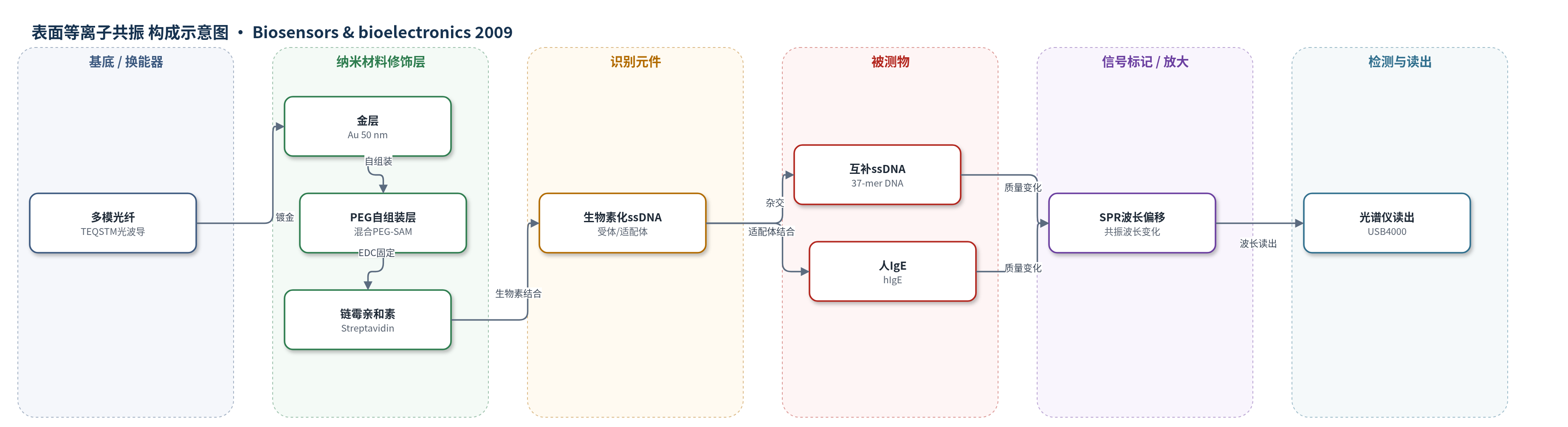
点击图片查看大图 · 依据论文自动绘制
传感器类型
表面等离子共振(SPR)生物传感器
检测对象
互补单链DNA(complementary ssDNA,37-mer)和人免疫球蛋白E(human immunoglobulin E, hIgE);样品基质为缓冲液(TRIS/NaCl、TGK),文中提出可拓展至人血清加标样品。
检测原理
该传感器基于光纤SPR的无标记实时检测原理。白光源经多模光纤传输至去包层金层表面,入射光激发金膜表面等离子体,反射光谱出现共振谷。当生物素化ssDNA或hIgE适配体与目标分子结合时,金表面附近的质量与折射率增加,SPR共振条件改变,共振波长向长波方向移动。表面采用混合PEG自组装单分子层抑制非特异性吸附,链霉亲和素通过EDC固定并捕获生物素化识别元件。DNA杂交或适配体–hIgE结合事件直接转化为SPR波长偏移,光谱仪实时记录偏移量;结合量越大,波长偏移越大。系统还可通过Langmuir模型拟合结合/解离曲线,获得动力学参数。
检测灵敏度
DNA杂交: 线性范围: 0.5–5 μM;hIgE: LOD: 2 ± 1 nM;LOQ: 6 ± 2 nM;线性范围: 6–100 nM;质量灵敏度: 0.0035 nm per pg/mm2;折射率分辨率: 2.10−4 RIU
效应效果
该传感器表现出良好的选择性与稳定性。非互补ssDNA和hIgG均不产生显著SPR偏移,说明PEG层有效抑制非特异性吸附。蔗糖重复测量平均变异系数为0.3%,不同光纤斜率差异小于5%。DNA涂层表面经酸处理可再生至少20次,性能无显著变化,仅出现轻微基线下漂。与Biacore 3000和毛细管电泳相比,光纤SPR测得hIgE适配体解离常数为30.9±2.9 nM,Biacore为20±2 nM,ACE为52±10 nM,结果相互印证。系统无需微流控,成本约为商业SPR平台的1/2,适合低成本、可重复的DNA与蛋白定量及结合动力学研究,作者认为可拓展至医疗诊断、环境检测和食品安全。
传感器的构成
- 基底/光波导:TEQSTM多模光纤(Thorlabs,400 μm,NA 0.39),去包层后作为SPR敏感区基底与光传输通道
- 金属换能层:50 nm金层(Au),溅射沉积,支持表面等离子体共振并反射光
- 抗非特异修饰层:混合PEG自组装单分子层(5% SHC11(PEO)6COOH + 95% SHC11(PEO)3OH),形成亲水抗蛋白吸附层
- 活化/交联剂:EDC(1-乙基-3-[3-二甲基氨基丙基]碳二亚胺盐酸盐),活化PEG羧基以共价固定链霉亲和素
- 结合元件:链霉亲和素(Streptavidin, SA),特异性结合生物素化ssDNA/适配体
- 识别元件:生物素化单链DNA(biotinylated ssDNA)或抗hIgE DNA适配体(hIgE aptamer),用于捕获互补ssDNA或hIgE
- 再生剂:稀盐酸(2 mM HCl或1.25 mM HCl),用于解链再生DNA杂交表面
中文摘要
本文报道了一种光纤表面等离子共振(SPR)生物传感器,用于无标记、可重复且低成本地检测DNA杂交和DNA–蛋白相互作用。该工作首次将光纤SPR系统与DNA适配体生物受体相结合。光纤表面溅射50 nm金层,并修饰由羧基和羟基封端聚乙二醇(PEG)巯基十一烷硫醇组成的混合自组装单分子层,以降低非特异性吸附;链霉亲和素通过EDC活化固定于PEG羧基端,用于捕获生物素化单链DNA(ssDNA)。在DNA–DNA杂交实验中,传感器可检测与表面受体ssDNA具有15个核苷酸重叠的37-mer ssDNA,在0.5–5 μM范围内呈线性响应,阴性对照无显著非特异性结合,且传感器可再生。在第二项实验中,传感器表面固定抗人免疫球蛋白E(hIgE)的ssDNA适配体,检出限为2 nM,定量限为6 nM。该传感器还可实时监测表面结合动力学,测得适配体与hIgE的解离常数为30.9±2.9 nM,与文献及对照实验一致。
英文摘要
In this paper we present a fiber optic surface plasmon resonance (SPR) sensor as a reusable, cost-effective and label free biosensor for measuring DNA hybridization and DNA-protein interactions. This is the first paper that combines the concept of a fiber-based SPR system with DNA aptamer bioreceptors. The fibers were sputtered with a 50nm gold layer which was then covered with a protein repulsive self-assembled monolayer of mixed polyethylene glycol (PEG). Streptavidin was attached to the PEG's carboxyl groups to serve as a versatile binding element for biotinylated ssDNA. The ssDNA coated SPR fibers were first evaluated as a nucleic acid biosensor through a DNA-DNA hybridization assay for a random 37-mer ssDNA. This single stranded DNA showed a 15 nucleotides overlap with the receptor ssDNA on the SPR fiber. A linear calibration curve was observed in 0.5-5 microM range. A negative control test did not reveal any significant non-specific binding, and the biosensor was easily regenerated. In a second assay the fiber optic SPR biosensor was functionalized with ssDNA aptamers against human immunoglobulin E. Limits of detection (2nM) and quantification (6nM) in the low nanomolar range were observed. The presented biosensor was not only useful for DNA and protein quantification purposes, but also to reveal the binding kinetics occurring at the sensor surface. The dissociation constant between aptamer and hIgE was equal to 30.9+/-2.9nM. The observed kinetics fully comply with most data from the literature and were also confirmed by own control measurements.