组成图示
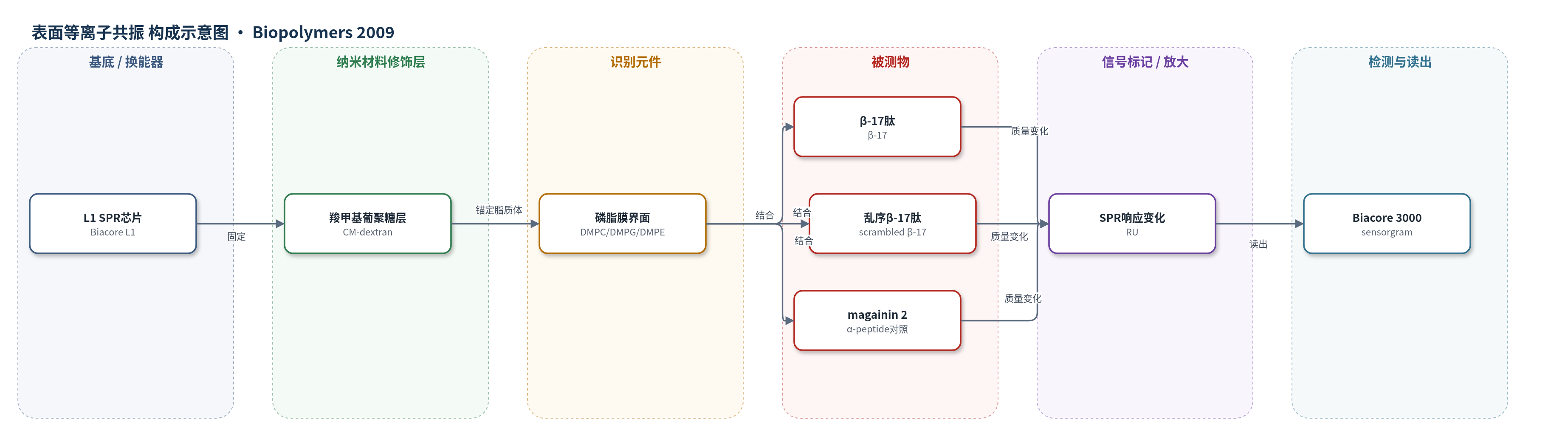
点击图片查看大图 · 依据论文自动绘制
传感器类型
表面等离子共振(SPR)生物传感器
检测对象
抗菌肽 β-17、乱序 β-17(scrambled β-17)、magainin 2;样品基质为磷脂脂质体膜模拟物(DMPC、DMPG、DMPE、cholesterol 混合物)及磷酸盐缓冲液
检测原理
SPR 检测中,L1 芯片表面的疏水基团将磷脂脂质体固定为膜界面。抗菌肽注入后,首先通过静电作用接近带负电或两性膜表面,并伴随二级结构诱导或保持;随后疏水残基驱动肽插入脂质双层。肽结合使界面质量与折射率增加,引起 SPR 共振角偏移,仪器以响应单位 RU 记录结合量。随着肽浓度升高,结合响应增大;低浓度时结合很少,高浓度时快速达到平衡。β-17 因两亲性可部分插入膜内,解离较慢,600 s 后仍残留约 20%–30% 结合;scrambled β-17 缺乏连续疏水面,几乎完全解离。该过程无酶或核酸放大,信号直接来自肽-膜结合质量变化。
检测灵敏度
原文未报告 LOD、线性范围、灵敏度斜率或相关系数。
效应效果
SPR 结果显示,β-17 与 scrambled β-17 在四种磷脂膜上的结合水平相近,且均明显高于 magainin 2;在 DMPC 上,12 μM 时 β-17 约 1500 RU,scrambled β-17 约 2000 RU,而 magainin 2 仅约 750 RU。结合量总体顺序为 DMPE/DMPG > DMPC ≈ DMPC/DMPG > DMPC/DMPG/cholesterol。β-17 解离较慢,600 s 后残留约 20%–30% 结合,scrambled β-17 几乎完全解离,含胆固醇膜上两者均快速解离。由于低浓度结合弱且存在阈值,β-肽传感器图难以用 Langmuir、parallel 或 two-state 模型良好拟合;magainin 2 拟合较好。作者认为活性差异主要源于插入程度和解离速率,而非结合量,为 β-肽抗菌剂设计提供依据。
传感器的构成
- 基底/换能器:Biacore L1 传感器芯片,SPR 换能表面,用于固定脂质膜并检测结合响应
- 修饰层:羧甲基葡聚糖(carboxymethylated dextran)及其共价连接的疏水基团,提供脂质体锚定位点
- 识别/界面层:磷脂小单层/脂质体膜(DMPC、DMPG、DMPE、cholesterol 混合物,SUV 50 nm),模拟哺乳动物或微生物膜
- 分析物:抗菌肽 β-17、乱序 β-17(scrambled β-17)和 magainin 2,作为被测结合分子
- 清洗/稳定剂:CHAPS 清洗液和 10 mM NaOH,用于去除多膜结构并稳定基线
- 读出系统:Biacore 3000 SPR 分析仪,输出传感器图响应单位 RU
中文摘要
本研究利用表面等离子共振(SPR)生物传感器考察两种β-肽与磷脂膜的相互作用。研究对象为新型β-氨基酸抗菌肽β-17及其非抗菌乱序类似物scrambled β-17,并以已知抗菌肽magainin 2作为对照。实验采用模拟哺乳动物细胞膜(富含磷脂酰胆碱和胆固醇)和微生物细胞膜(富含磷脂酰乙醇胺和磷脂酰甘油)的多种磷脂混合物,构建脂质体膜界面。SPR用于测定肽在不同膜上的结合量、结合速率和解离行为;圆二色性(CD)用于分析肽在水相缓冲液及脂质体存在下的二级结构。结果显示,β-17与scrambled β-17长度、电荷相同,在水相和脂质体中均具有相似二级结构,并在各膜上结合水平相近,说明二者生物活性差异并非主要由结合量决定,而与插入程度和膜解离速率有关。两种β-肽在所有膜上的结合均高于magainin 2,提示肽的两亲性与二级结构诱导在膜结合中共同起重要作用。
英文摘要
The membrane interaction of two beta peptides was studied using a surface plasmon resonance biosensor. The two peptides are beta-17, a novel beta-amino acid based antimicrobial peptide and the corresponding scrambled-beta17--a non-antimicrobial beta-peptide analogue. Membrane interaction studies were performed with a series of phospholipid mixtures which mimic either mammalian cells (high in phosphatidylcholine and cholesterol) or microbial cells (high in phosphatidylethanolamine and phosphatidylglycerol). The results were compared with the membrane binding of the well-characterized antimicrobial peptide magainin 2. The secondary structure of these peptides were also determined in each lipid mixture by circular dichroism and correlated with the membrane-binding properties. Both beta-17 and the scrambled peptide have the same peptide length, charge and showed a similar secondary structure in both aqueous buffer and in the presence of liposomes. Both peptides also bound to a similar level on each membrane mixture, showing that the dramatic difference in biological activity is not based on the amount of peptide bound but rather differences in the degree of insertion and rate of membrane dissociation. Although beta-17 and the scrambled beta-17 peptide exhibited similar binding properties on all membrane mimics, both beta-peptides bound more to all membranes compared with magainin 2. Overall, the results further reveal the significant interplay between peptide amphipathicity and secondary structure induction on membrane binding.