组成图示
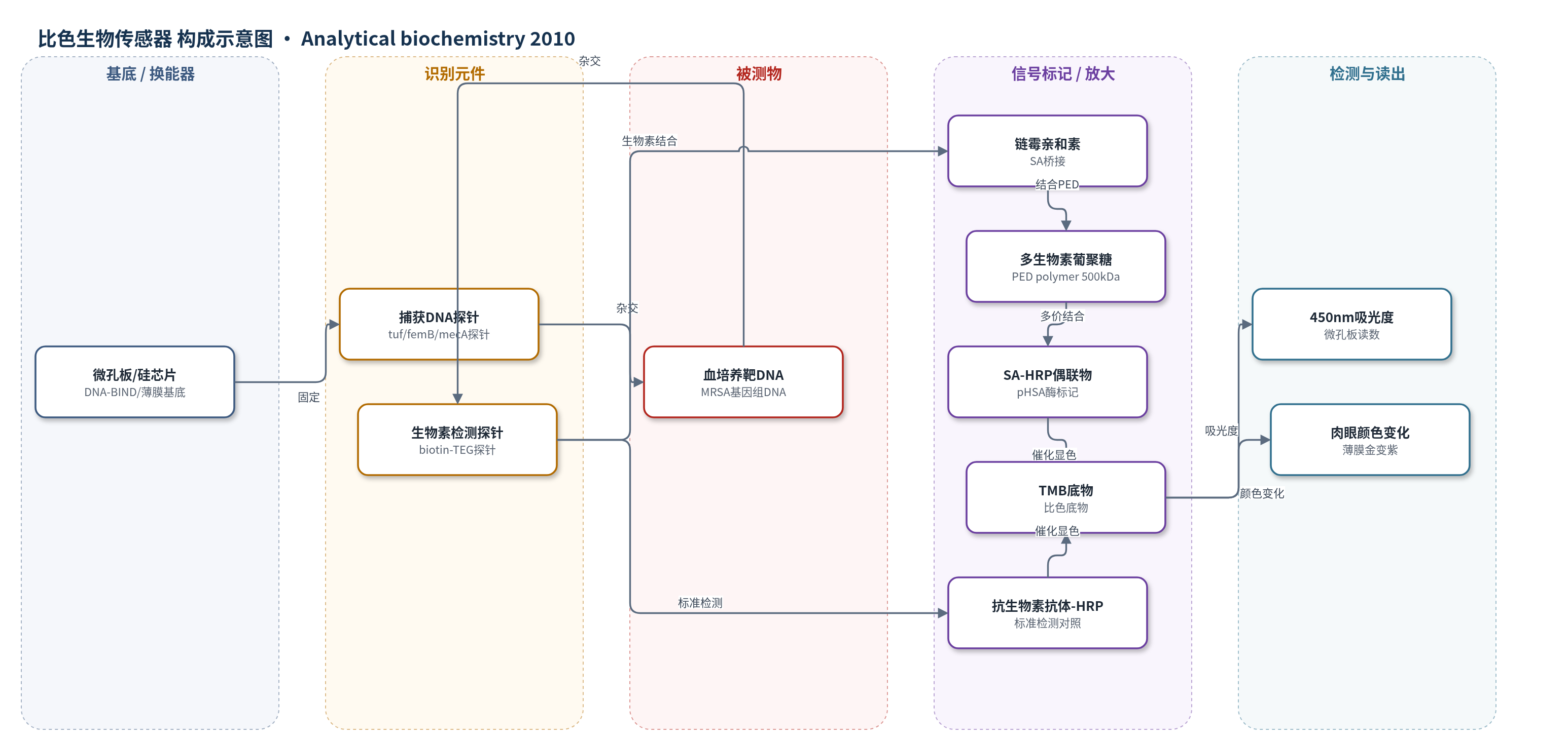
点击图片查看大图 · 依据论文自动绘制
传感器类型
比色生物传感器
检测对象
葡萄球菌属基因组DNA(Staphylococcus genus DNA, tuf)、金黄色葡萄球菌基因组DNA(Staphylococcus aureus DNA, femB)、耐甲氧西林金黄色葡萄球菌(MRSA)mecA基因;样品基质:阳性血培养瓶(positive blood culture bottles)
检测原理
表面固定 DNA 捕获探针与阳性血培养裂解液中的靶基因组 DNA 杂交,随后生物素标记检测探针与靶 DNA 杂交,形成表面固定三联复合物。PED 放大:链霉亲和素(SA)结合检测探针末端生物素,500 kDa 多生物素化葡聚糖(PED polymer,约80 biotin/monomer)结合 SA,形成高密度生物素界面;再结合 SA-HRP 偶联物,使每个杂交事件招募多个 HRP。HRP 催化 TMB 氧化显色,产物在微孔板中产生 450 nm 吸光度,或在薄膜芯片上形成沉淀使颜色由金变紫。靶 DNA 浓度越高,表面杂交复合物越多,招募的 HRP 越多,显色信号越强。无需 PCR 靶标扩增,依靠多价生物素-链霉亲和素相互作用实现信号放大。
检测灵敏度
LOD: limits of detection as low as 1 fmol/L target DNA;LOD: 120 to 300 fM target sequence;LOD: 4 and 1 fM for femB and tuf;LOD: 0.5 to 2 × 10^6 CFU/ml;标准检测 LOD: 111 fM;标准检测 LOD: ≈10 pM
效应效果
PED 在模型杂交中无显著非特异背景,检出限提高 10–100 倍;微孔板法 8 min 杂交即可见显著信号,15 min 饱和,总检测时间 35 min,而标准法 30 min 仍无显著信号。薄膜生物传感器标准法肉眼 LOD 为 111 fM,PED 将 femB 和 tuf 的 LOD 分别降至 4 fM 和 1 fM,提高约 25–100 倍。对 MRSA 阳性血培养,标准法 45 min 才出现弱信号,PED 在 10 min 内可见、5 min 后明确,总时间 25–30 min;连续混合使信号提高约 3 倍。与实时 PCR 约 2 h 相比,作者认为其更快、成本低于 0.01 美元/测试且无 PCR 污染风险。未报告 RSD、回收率或长期稳定性。
传感器的构成
- 基底/换能器:微孔板 DNA-BIND 表面或硅薄膜生物传感器芯片(thin film biosensor),用于固定捕获探针并承载杂交与显色
- 识别元件:DNA 捕获探针(tuf/femB/mecA 基因特异性),5′-I-linker 和 S-18 spacer 用于表面连接
- 识别元件:生物素标记 DNA 检测探针(biotin-TEG,X),与靶 DNA 杂交形成三联复合物
- 信号桥接:链霉亲和素(SA),结合表面生物素并提供多价结合位点
- 信号放大层:500 kDa 氨基葡聚糖多生物素化聚合物(PED polymer,约80 biotin/monomer),结合 SA 放大生物素数量
- 酶标记物:链霉亲和素-辣根过氧化物酶偶联物(SA-HRP 或 poly-HRP SA, pHSA),催化 TMB 显色
- 底物/显色剂:四甲基联苯胺(TMB),HRP 催化产生比色信号;微孔板用可溶性 Super Slow-HRP TMB,薄膜用沉淀 TMB
- 封闭剂:无生物素酪蛋白(biotin-free casein)封闭微孔板;杂交缓冲液含碱处理酪蛋白和 Tween 20
中文摘要
本文描述一种简单廉价的信号放大方法,称为聚合物酶检测(PED),可快速灵敏地直接从阳性血培养瓶中检测识别葡萄球菌属的 tuf 基因保守序列、特异性检测金黄色葡萄球菌的 femB 基因保守序列以及耐甲氧西林基因 mecA。将微生物特异性捕获探针固定于微孔板或硅芯片上;靶序列与生物素标记的靶特异性探针杂交到互补捕获探针上,形成表面固定的生物素标记三联复合物。通过两步过程放大信号:先以链霉亲和素(SA)孵育表面固定生物素,再加入约携带 80 个生物素的 500 kDa 葡聚糖聚合物。随后结合 SA-辣根过氧化物酶(HRP)偶联物,并用四甲基联苯胺(TMB)显色。PED 使模型 DNA 杂交检测的检出下限提高 10–100 倍,检出限低至 1 fmol/L 靶 DNA。该灵敏度使利用薄膜生物传感器芯片或微孔板法在 25–35 min 内检测耐甲氧西林金黄色葡萄球菌阳性血培养中的基因组 DNA 成为可能。
英文摘要
This article describes a simple and inexpensive signal amplification method, termed polymeric enzyme detection (PED), which permits rapid and sensitive detection of conserved sequences in the tuf gene that identify Staphylococcus genus, conserved sequences in the femB gene that specifically detect Staphylococcus aureus species, and the methicillin resistance gene mecA directly from positive blood culture bottles. Microbe-specific capture probes were immobilized onto microtiter plates or silicon chips. Target sequences and biotin-labeled, target-specific probes were hybridized to complementary capture probes to create a biotin-labeled, surface-immobilized tripartite complex. In a two-step process, signal was amplified by incubating the surface-immobilized biotin with streptavidin followed by the addition of a 500-kDa dextran polymer conjugated with approximately 80 biotins. Signal was then developed by binding of a streptavidin-horseradish peroxidase conjugate followed by incubation with the substrate tetramethylbenzidine. Use of the PED method improved the lower limit of detection 10- to 100-fold in model DNA hybridization assays with limits of detection as low as 1 fmol/L target DNA. This level of sensitivity permits detection of genomic DNA from methicillin-resistant S. aureus positive blood cultures within 25 to 35 min using either a thin film biosensor chip or a microtiter plate-based assay.