组成图示
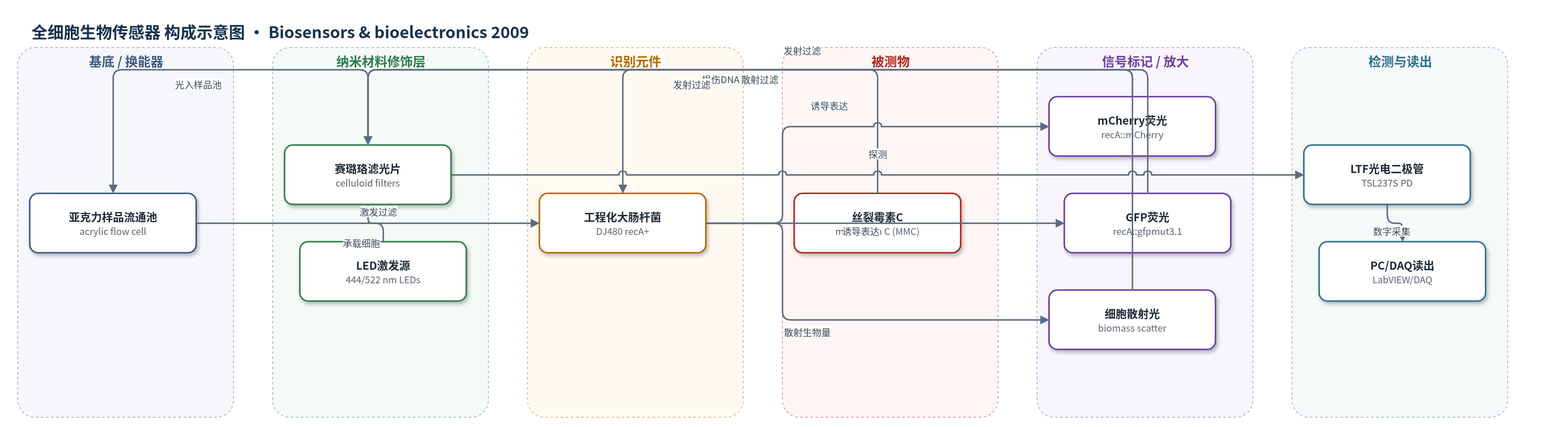
点击图片查看大图 · 依据论文自动绘制
传感器类型
全细胞生物传感器
检测对象
丝裂霉素 C(mitomycin C, MMC);样品基质:大肠杆菌培养液/液体悬浮液(Neidhardt EZ-Rich 培养基)
检测原理
丝裂霉素 C(MMC)进入大肠杆菌后造成 DNA 损伤,修复过程产生单链 DNA,激活 SOS 应答中的 RecA 蛋白,使 recA 启动子转录荧光报告基因。mCherry 或 GFPmut3.1 随 MMC 浓度升高而积累,形成可检测荧光。流通池中的 444 nm 或 522 nm LED 经赛璐珞滤光片激发样品,发射荧光与 90° 散射光由带滤光片的 LTF 光电二极管接收,并转换为数字脉冲频率。系统同时读取荧光通道和生物量散射通道,用双方程响应面校准估计未稀释样品中的荧光蛋白比例与细胞密度,从而消除高密度自屏蔽和内滤效应。信号放大主要来自 SOS 转录诱导和荧光蛋白长时间积累。
检测灵敏度
LOD: 0.25 nM(recA::mCherry,SNR=3);2.0 nM(recA::gfpmut3.1,SNR=3);11.7 nM(recA::mCherry,2倍诱导);>100 nM(recA::gfpmut3.1,2倍诱导)
效应效果
在该 LED/LTF 流通荧光仪中,recA::mCherry 的 MMC 响应覆盖约 4 个数量级,峰值诱导因子约 5;recA::gfpmut3.1 仅约 3 个数量级,峰值约 2。mCherry 在高细胞密度下光学失真明显小于 GFP,GFP 受自屏蔽和内滤效应影响更大。双方程校准可在 OD620 约 0.1–2.1 的未稀释样品中同时估计荧光比例与生物量。与商业微孔板分光光度计相比,LTF 系统在高细胞密度下对 GFP 表现相当,对 RFP 某些情况更好。使用主曲线时,4 个月内仍可可靠估计约 10 nM MMC。作者认为该平台低成本、紧凑、适合多通道、分布式或可丢弃全细胞传感。
传感器的构成
- 样品流通池:透明亚克力(acrylic)样品室,约3 mm×4 mm×8 mm、体积约95 µL,承载细胞悬液并形成光路。
- 光学激发源:高亮 LED,444 nm 蓝 LED 与 522 nm 绿 LED,交替脉冲激发荧光和散射。
- 滤光片层:赛璐珞滤光片(Rosco #346、#12、#389、#26),置于激发与发射光路,选择波长并降低背景。
- 生物识别元件:工程化大肠杆菌 DJ480(E. coli MG1655 Δ(argF-lac)U169,lacI− recA+),作为全细胞识别元件。
- 报告基因/信号标记:recA::mCherry 或 recA::gfpmut3.1 融合蛋白,DNA 损伤诱导 SOS 响应后表达荧光。
- 质粒载体:pTrc99a 衍生中等拷贝质粒,含 recA 启动子、MCS、rrnBT1T2 终止子、氨苄抗性、pBR322 ori。
- 检测器:光频转换硅光电二极管 LTF PD(TSL237S,TAOS),将光信号转为数字脉冲频率。
- 控制与读出:PC、LabVIEW 与 NI DAQcard 6024E,管理 LED 脉冲、选择通道并记录频率信号。
中文摘要
本研究开发了一种微型流通式光学检测池,有望集成为独立、可丢弃的全细胞生物传感器平台。该紧凑低成本光学系统由紧密耦合的发光二极管(LED)、光频转换(LTF)光电二极管和赛璐珞滤光片组成,用于测量液体悬浮液中生物传感器细胞产生的荧光报告子。以携带中等拷贝质粒、荧光报告基因融合于 recA 启动子的大肠杆菌为演示对象,细胞暴露于 DNA 损伤剂丝裂霉素 C(MMC)。比较绿色荧光蛋白(GFPmut3.1)与红色荧光蛋白(mCherry)后,mCherry 在两方面优于 GFP:高密度样品引起的光学失真更小,且 MMC 检出限更低,recA::mCherry 估计为 0.25 nM,recA::gfpmut3.1 为 2.0 nM。此外,提出多通道光学数据集成校准方法,可同时测量荧光和生物量浓度,显著消除浓样品光学失真,避免测量前稀释。
英文摘要
A miniature flow-through optical cell has been developed with the potential for integration into a stand-alone, potentially disposable whole-cell biosensor platform. The compact and inexpensive optical system is comprised of closely coupled light-emitting diodes (LEDs), light-to-frequency (LTF) photodiodes, and celluloid filters. The system has been optimized to measure fluorescent reporters produced by cultures of biosensor cells in liquid suspension. As demonstration subjects, Escherichia coli cells carrying medium-copy plasmids with fluorescent reporter fusions to the rec promoter were exposed to the DNA-damaging agent mitomycin C (MMC). As reporter proteins, green fluorescent protein (GFP) and red fluorescent protein (RFP) were compared for suitability in the compact instrument. The RFP mCherry outperformed GFP (GFPmut3.1) as a reporter protein in the developed system on two counts. First, measurement distortions due to high optical density suspensions are minimal using RFP compared to GFP. Second, the limit of detection for MMC is estimated at 0.25nM for recA::mCherry and 2.0nM for recA::gfpmut3.1. Finally, a measurement method is presented whereby multiple channels of optical data are calibrated in an integrated fashion to allow simultaneous measurement of fluorescence and biomass concentration. The method substantially eliminates optical distortions due to dense samples and thus obviates the conventional need for sample dilution prior to measurement.