组成图示
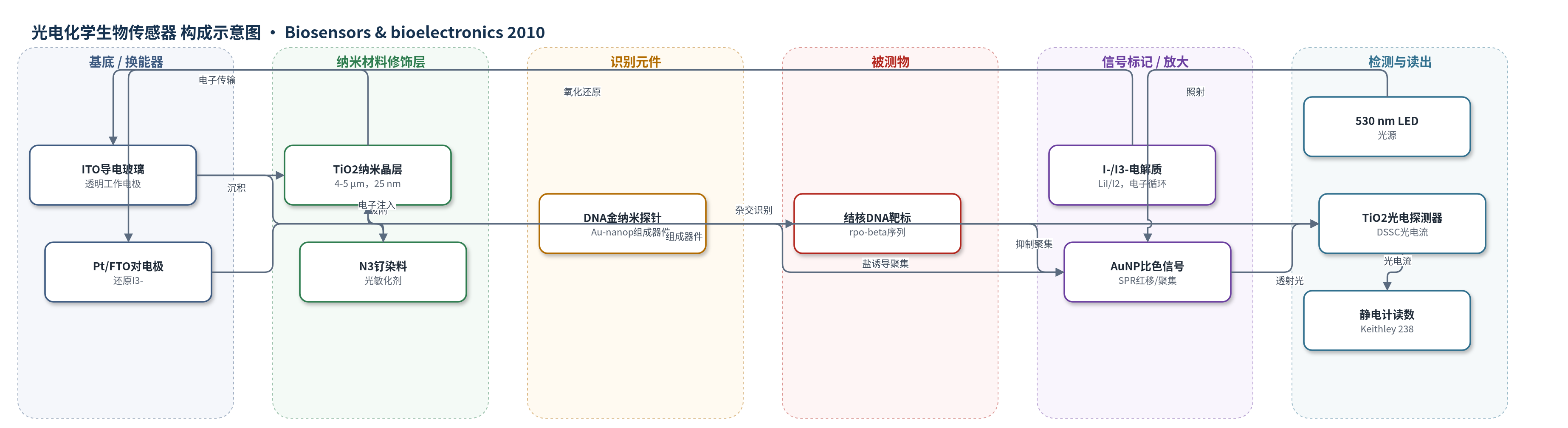
点击图片查看大图 · 依据论文自动绘制
传感器类型
光电化学生物传感器
检测对象
结核分枝杆菌rpo-beta基因特异性DNA序列(Mycobacterium tuberculosis rpo-beta DNA);样品基质:磷酸盐缓冲液中的DNA寡核苷酸(非临床样品)
检测原理
该传感器以DNA功能化金纳米探针为识别元件。Au-nanoprobe与互补结核DNA靶标杂交后,加入NaCl/MgCl2时盐诱导的聚集被抑制,金纳米颗粒保持分散,SPR峰位于约520 nm,溶液呈红色;非互补靶标不能杂交,金纳米颗粒聚集,SPR红移,溶液变蓝,530 nm附近吸光增强。530 nm LED照射样品池,透射光进入染料敏化TiO2光电探测器:N3染料吸收光子后电子注入TiO2导带,经ITO外电路流向Pt/FTO对电极,I-在电解质中还原I3-,形成光电流。检测响应Rdet为参考溶液与样品溶液光电流密度之差。金纳米颗粒聚集程度和浓度改变透射光强,使光电流随被测DNA是否互补及AuNP浓度变化而变化。
检测灵敏度
LOD: 1.0 nM(doctor blade);LOD: 1.5 nM(inkjet printed);R^2 > 0.98
效应效果
该传感器对互补与非互补DNA靶标具有明显区分能力:DBP光电探测器检测响应差异为17.5%,IPP为6.7%;非互补体系中AuNP聚集使SPR最大位移26 nm。与分光光度法相比,DBP灵敏度接近(LOD 1.0 nM,分光光度计优于0.5 nM),IPP为1.5 nM,作者认为两者与1.0 nM比色法相当。测量重复6次,结果一致且可重现,但因LED半宽约30–35 nm及系统噪声,波动大于分光光度计。研究未使用临床样品,仅在缓冲液DNA寡核苷酸中验证。作者强调喷墨打印可降低成本、实现一次性光电探测器,适合发展中国家分子诊断,并计划进一步微流控集成。
传感器的构成
- 基底/工作电极:ITO导电玻璃(indium doped tin oxide, ITO, 10–12 Ω sq−1),提供透明导电基底与电子收集。
- 活性层:TiO2纳米晶薄膜(Solaronix TiO2分散液,粒径约25 nm,喷墨厚度4–5 μm或刮刀约7 μm),作为光阳极和电子传输层。
- 敏化层:N3钌染料(cis-di(thiocyanato)bis(2,2-bipyridyl-4,4-dicarboxylate) ruthenium(II)),吸附于TiO2表面,吸收530 nm附近光并注入电子。
- 对电极:Pt/FTO导电玻璃(platinum-coated fluorine doped tin oxide, Pt/FTO),作为对电极并催化I3-还原。
- 电解质:0.5 M LiI与0.05 M I2的乙腈/碳酸丙烯酯(6:4, v/v)溶液,提供I-/I3-氧化还原循环。
- 识别元件:DNA功能化金纳米探针(Au-nanoprobes,AuNPs修饰巯基寡核苷酸5′-thiol-GGACGTGGAGGCGATC-3′),识别M. tuberculosis rpo-beta序列。
- 信号标记/比色元件:金纳米颗粒(AuNPs)表面等离子共振(SPR)颜色变化,互补靶标抑制聚集保持红色,非互补靶标盐诱导聚集变蓝。
- 样品池:10 mm光程塑料比色皿,盛装AuNP/Au-nanoprobe溶液并置于LED与探测器之间。
中文摘要
本文报道了一种将染料敏化TiO2光电探测器与基于DNA功能化金纳米颗粒的非交联杂交比色法相结合的一次性DNA生物传感器。作者采用喷墨打印技术制备染料敏化TiO2光电探测器,这是一种非接触、数字、无掩模、无真空的增材制造方法,有利于低成本批量生产,并与传统刮刀法制备的光电探测器进行了比较。该传感器利用DNA修饰的金纳米探针识别结核分枝杆菌rpo-beta基因中的特定序列:加入盐后,非互补靶标不能阻止金纳米颗粒聚集,溶液由红变蓝;互补靶标则抑制聚集,保持红色。530 nm LED照射样品,透射光被染料敏化TiO2光电探测器转换为光电流。刮刀法系统可检测低至1.0 nM的金纳米颗粒聚集,喷墨打印系统为1.5 nM,灵敏度与分光光度法相当。对互补与非互补金纳米探针检测,传统光电探测器响应差异为17.5%,喷墨打印为6.7%。该平台有望降低分子诊断成本,尤其适用于发展中国家。
英文摘要
A dye sensitized TiO(2) photodetector has been integrated with a DNA detection method based on non-cross-linking hybridization of DNA-functionalized gold nanoparticles, resulting in a disposable colorimetric biosensor. We present a new approach for the fabrication of dye sensitized TiO(2) photodetectors by an inkjet printing technique-a non-contact digital, additive, no mask and no vacuum patterning method, ideal for cost efficient mass production. The developed biosensor was compared against a dye sensitized photodetector fabricated by the traditional "doctor blade" method. Detection of gold nanoparticle aggregation was possible for concentrations as low as 1.0 nM for the "doctor blade" system, and 1.5 nM for the inkjet printed photodetector. The demonstrated sensitivity limits of developed biosensors are comparable to those of spectrophotometric techniques (1.0 nM). Our results show that a difference higher than 17% by traditional photodetector and 6% by inkjet printed in the photoresponses for the complementary and non-complementary gold nanoprobe assays could be attained for a specific DNA sequence from Mycobacterium tuberculosis, the etiologic agent of human tuberculosis. The decrease of costs associated with molecular diagnostic provided by a platform such as the one presented here may prove of paramount importance in developing countries.