组成图示
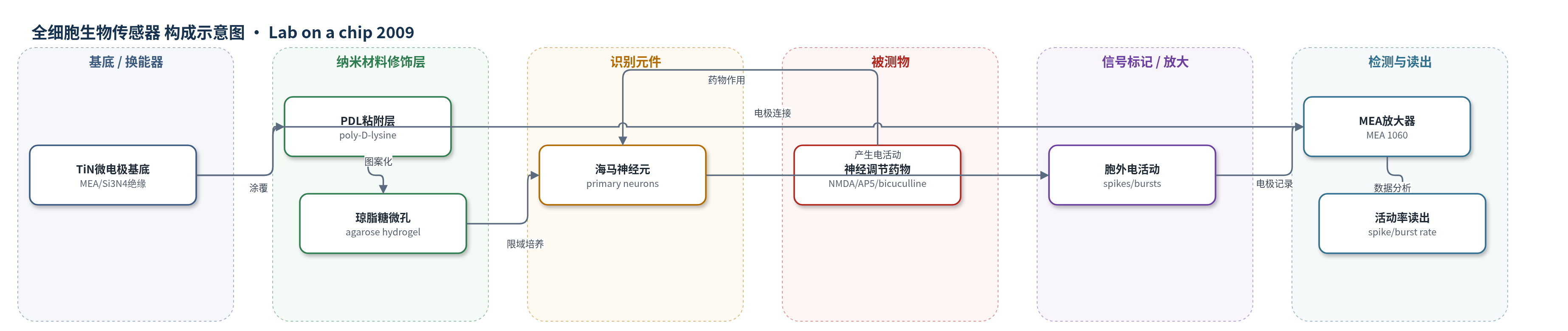
点击图片查看大图 · 依据论文自动绘制
传感器类型
全细胞生物传感器
检测对象
NMDA(N-methyl-D-aspartic acid)、AP5(NMDA受体拮抗剂)、bicuculline(bicuculline),样品基质为神经元培养液(Neurobasal medium)
检测原理
该传感器以琼脂糖微孔限域培养的原代海马神经元作为识别与响应元件。药物分子进入培养液后,与神经元突触受体发生特异性作用:NMDA激动兴奋性谷氨酸受体,AP5阻断NMDA受体,bicuculline阻断抑制性GABA受体。受体状态改变引起神经元膜电位和突触传递变化,使单个微孔内神经元网络产生或抑制胞外动作电位,表现为spike rate和burst rate变化。TiN微电极通过电容耦合记录胞外电位,MEA放大器放大并数字化,软件检测阈值以上尖峰并计算活动率。琼脂糖微孔将神经元限制在约100 μm区域,提高局部神经元密度至约1400 cell/mm2,加速网络成熟并增强药物响应,同时隔离各微电路,实现单芯片多通道并行读出。
检测灵敏度
—
效应效果
平台在培养液中稳定超过3周,干燥可存储1周;每cm2最多2500孔,微孔直径100 μm,水合厚度约13.3 μm。1 DIV时99%以上微孔至少含1个神经元,平均11.4±6.8个/孔。7 DIV活动微孔比例66.4±20.6%,burst微孔比例51.1±21.6%;平均spike幅度152 mVpp,范围30–697 mVpp。NMDA 20/40 μM增加spike rate;AP5 25/50 μM基本消除spike activity且可逆;bicuculline 20/40 μM使活动转为规律burst。微电路间cross-correlation近零,可替代重复MEA试验用于高通量药物筛选。
传感器的构成
- 基底/换能器电极:平面微电极阵列(MEA),含59个TiN微电极(直径30 μm、间距200 μm)和Si3N4绝缘层,用于记录胞外动作电位。
- 细胞粘附层:聚-D-赖氨酸(PDL,MW 70,000–150,000)涂覆于MEA表面,提供神经元粘附位点。
- 抗粘附隔离层:琼脂糖水凝胶(agarose hydrogel,2% w/v,凝胶强度>12000 g/cm2)覆盖非粘附区,形成微孔壁并隔离神经元微电路。
- 识别/响应元件:原代大鼠海马神经元(primary hippocampal neurons),在微孔内形成神经元微电路,作为生物识别与响应元件。
- 培养介质:Neurobasal培养基,添加B-27、Glutamax、L-glutamic acid和Penicillin-Streptomycin,维持神经元存活与电活动。
- 读出系统:MEA 1060放大器(增益1200、带宽10 Hz–5 kHz)、MC Rack数据采集(40 kHz)和NeuroExplorer分析,输出spike rate与burst rate。
中文摘要
对于基于细胞的生物传感器应用,解离神经元通常培养在平面微电极阵列(MEA)上,以测量网络电活动。为缩短数据采集时间并提高统计分析效力,需要多微孔型平台。本研究提出一种新方法,将传统MEA转换为多微孔MEA,并在其上构建微米级神经元培养阵列,即神经元微电路阵列。首先,在MEA表面涂覆细胞粘附层聚-D-赖氨酸(PDL),随后用细胞排斥层琼脂糖水凝胶进行图案化,既限定细胞粘附区域,又将各神经元微电路相互隔离。原代海马神经元在琼脂糖微孔MEA上培养数周,其自发电活动通过胞外动作电位进行表征。利用神经传递调节剂,还演示了同时监测多个神经元微电路的药物响应。该方法可用于需要重复试验以获得单个数据点的神经生物学功能分析或神经元生物传感器领域。
英文摘要
For cell-based biosensor applications, dissociated neurons have been cultured on planar microelectrode arrays (MEAs) to measure the network activity with substrate-embedded microelectrodes. There has been a need for a multi-well type platform to reduce the data collection time and increase the statistical power for data analysis. This study presents a novel method to convert a conventional MEA into a multi-well MEA with an array of micrometre-sized neuronal culture ('neuronal micro-circuit array'). An MEA was coated first with cell-adhesive layer (poly-D-lysine) which was subsequently patterned with a cell-repulsive layer (agarose hydrogel) to both pattern the cell adhesive region and isolate neuronal micro-circuits from each other. For a few weeks, primary hippocampal neurons were cultured on the agarose microwell MEA and the development of spontaneous electrical activities were characterized with extracellular action potentials. Using neurotransmission modulators, the simultaneous monitoring of drug responses from neuronal micro-circuit arrays was also demonstrated. The proposed approach will be powerful for neurobiological functional assay studies or neuron-based biosensor fields which require repeated trials to obtain a single data point due to biological variations.