组成图示
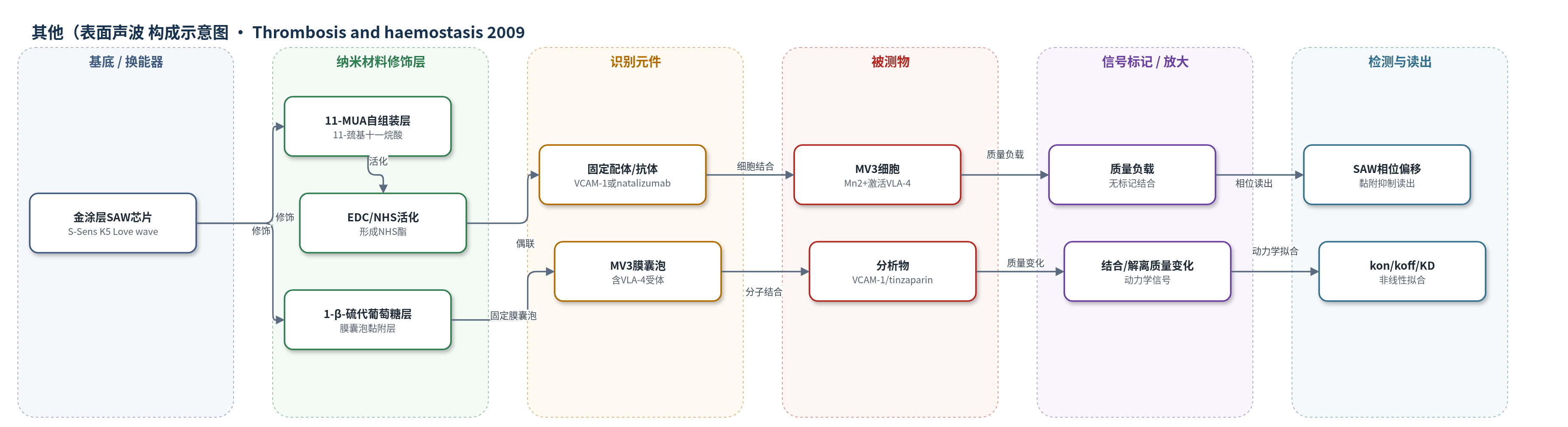
点击图片查看大图 · 依据论文自动绘制
传感器类型
其他(表面声波(SAW)生物传感器)
检测对象
替扎帕林(tinzaparin)、未分馏肝素(UFH)、fondaparinux、VCAM-1、MV3黑色素瘤细胞;样品基质为PBS缓冲液、细胞悬液和膜囊泡体系。
检测原理
SAW生物传感器基于压电Love wave,传感器表面质量负载改变声波相位。金芯片经11-MUA/EDC/NHS固定VCAM-1或natalizumab,或经1-β-硫代葡萄糖固定含VLA-4的MV3膜囊泡。注入VCAM-1、tinzaparin或Mn2+激活的MV3细胞后,识别事件在界面形成质量负载,相位上升;冲洗后解离使相位回落。结合速率随分析物浓度变化,按kobs=kon c+koff拟合得到kon、koff和KD。tinzaparin与VLA-4结合后通过空间位阻或构象变化干扰VCAM-1/细胞结合,从而抑制黏附。该体系无标记、无酶放大,直接读出分子相互作用。
检测灵敏度
未报告LOD、线性范围、灵敏度斜率或R^2;报告结合常数:VCAM-1 KD=1.01×10⁻⁸ M、kon=3.53×10^5 M-1s-1、koff=3.44×10⁻³ s-1;tinzaparin KD=4.61×10⁻⁶ M、kon=1.27×10^3 M-1s-1、koff=5.13×10⁻³ s-1;fondaparinux n.d.
效应效果
流动室中Mn2+使MV3黏附率由76.5%±6.9%升至87.3%±5.8%,natalizumab降至76.0%±4.2%;UFH2和tinzaparin在P-selectin阻断下分别降至74.3%±11.6%、72.5%±9.7%,fondaparinux无效。固定VCAM-1体系中UFH抑制强于tinzaparin,14–18单糖片段活性略降。SAW中tinzaparin显著抑制MV3与VCAM-1及natalizumab结合,1×10–9–1×10⁻⁴ M未见交叉结合。动力学:VCAM-1 KD=1.01×10⁻⁸ M,tinzaparin KD=4.61×10⁻⁶ M,koff接近,n>3。作者认为VLA-4是肝素抗转移新靶点。
传感器的构成
- 基底/换能器:金涂层SAW传感器芯片(S-Sens K5,Love wave),压电换能,将表面质量变化转为相位偏移
- 修饰层A:11-巯基十一烷酸(11-MUA)自组装单层,硫端结合金面,羧基端用于EDC/NHS偶联
- 识别元件A:重组人VCAM-1 Fc或抗VLA-4单抗natalizumab,固定于SAM上用于捕获/识别MV3细胞
- 封闭剂:1 M乙醇胺(pH 8.5),封闭未反应NHS酯,减少非特异结合
- 修饰层B:1-β-硫代葡萄糖(1-β-thioglucose)黏附层,用于原位固定MV3细胞膜囊泡
- 识别元件B:MV3黑色素瘤细胞膜囊泡(含VLA-4异二聚体),作为VLA-4受体识别元件
- 信号标记物:无标记(label-free),VCAM-1、tinzaparin或MV3细胞结合产生质量负载
- 读出层:SAW相位偏移随时间变化,经非线性拟合获得kon、koff和KD
中文摘要
肝素除抗凝外还具有抗转移作用,其机制可能涉及干扰黏附受体功能。本研究考察不同肝素对人黑色素瘤MV3细胞经VLA-4与固定VCAM-1黏附的抑制作用。未分馏肝素(UFH)和低分子肝素替扎帕林(tinzaparin)显示较强抑制活性;将tinzaparin分馏为14–18个单糖片段后抑制作用略降,而五糖fondaparinux无抑制效果,提示肝素分子大小影响VLA-4阻断。为在分子水平确认tinzaparin与VLA-4的结合,作者采用表面声波(SAW)生物传感器,将含VLA-4的MV3细胞膜囊泡固定于传感器表面,检测tinzaparin与VCAM-1的结合动力学。结果显示tinzaparin以低微摩尔亲和力结合VLA-4(KD=4.61×10⁻⁶ M),其解离常数与VCAM-1接近(5.13×10⁻³ s–1对3.44×10⁻³ s–1),表明存在特异性分子识别。该结果直接证实肝素可结合VLA-4,有助于理解肝素的抗转移活性。
英文摘要
Heparin possesses antimetastatic effects that were related to various molecular mechanisms beyond anticoagulant activities. The ability of heparin to interfere with the function of adhesion receptors in the metastatic course appears as a promising therapeutic approach. This refers to numerous findings that heparin attenuates metastasis in a selectin-dependent manner. We recently demonstrated that heparin interferes with the integrin VLA-4 on murine melanoma cells binding to VCAM-1. To confirm this activity and to obtain further insight into molecular recognition of heparin by VLA-4, we investigated the inhibition of VLA-4 mediated binding of human melanoma MV3 cells to immobilised VCAM-1 by different heparins. The size of heparin has an important impact on inhibition. Unfractionated heparin (UFH) and tinzaparin, a low-molecular-weight heparin (LMWH) representing a mean of about 18-20 monomers, displayed high inhibitory activity. Fractionating tinzaparin to 14-18 monomers reduced inhibition slightly, while the pentasaccharide fondaparinux was without effects. To confirm molecular recognition of tinzaparin by VLA-4, a surface acoustic wave-biosensor was applied. A VLA-4 containing membrane preparation of MV3 cells was immobilised at the sensors to allow for detection of kinetic binding constants of tinzaparin compared to VCAM-1. Tinzaparin binds to VLA-4 with affinity in the low micromolar range (4.61 x 10(-6) M), which clearly indicates specific molecular recognition. Furthermore, tinzaparin displays a nearly identical k(off) compared to VCAM-1 (5.13 x 10(-3) s(-1) versus 3.44 x 10(-3) s(-1)) which is evident for interference with the ligand binding. The data provide evidence for a direct confirmation of heparin binding to VLA-4 and thus, contribute to understand the antimetastatic activity of heparin.