组成图示
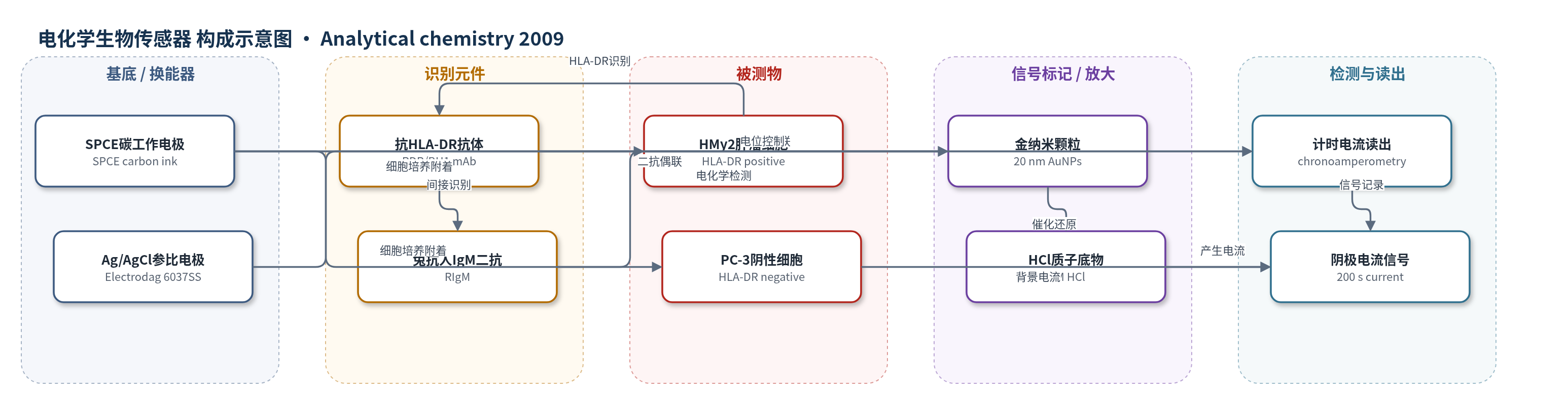
点击图片查看大图 · 依据论文自动绘制
传感器类型
电化学生物传感器
检测对象
肿瘤细胞(tumor cells,HMy2 HLA-DR 阳性 B 细胞系、PC-3 HLA-DR 阴性前列腺癌细胞系);样品基质为细胞悬液/培养液(700 µL,RPMI 1640 含 10% FCS)
检测原理
肿瘤细胞在 SPCE 工作区表面培养并附着,细胞表面 HLA-DR 与抗 HLA-DR 抗体结合;直接法使用 AuNP/RDR,间接法先结合 BH1 一抗,再用 AuNP/RIgM 二抗,使每个一抗可结合多个 AuNP,实现信号放大。未结合抗体用 PBS 洗去。加入 1 M HCl 后,在 -1.00 V 下 AuNPs 催化 H+ 还原为 H2,产生阴极电流;电流大小与电极表面 AuNP 数量成正比,进而与 HLA-DR 阳性细胞数量成正比。计时电流法记录 200 s 电流作为分析信号。无 AuNP 的 PC-3 阴性细胞仅产生背景电流。
检测灵敏度
LOD: 4000 cells per 700 µL;线性范围: 10000–200000 cells;灵敏度斜率: 0.0641 µA/(cell number/1000);R^2 = 0.9955
效应效果
该传感器对 HLA-DR 阳性 HMy2 细胞产生明显电催化信号,而对 HLA-DR 阴性 PC-3 细胞无信号,显示良好选择性;使用识别两种细胞的 32.4 mAb 作阳性对照时,PC-3 信号高于 HMy2,与流式细胞术免疫荧光结果一致。在 HMy2/PC-3 混合体系中,PC-3 不显著干扰 HMy2 信号。方法重现性 RSD 为 7%(100000 细胞,n=3),未报告实际临床样品加标回收率。最终检测仅需数分钟,无需昂贵仪器和复杂化学试剂,成本低于流式细胞术等现有方法,适合微型化、一次性使用和床旁诊断,可拓展至肿瘤转移、炎症细胞及蛋白质/DNA 检测。
传感器的构成
- 基底/换能器电极:丝网印刷碳电极(SPCE),碳工作电极(Electrodag 423SS carbon ink)、银/氯化银参比/辅助电极(Electrodag 6037SS silver/silver chloride ink)和绝缘层(Minico 7000 Blue),提供细胞附着与电化学检测界面。
- 识别元件:抗 HLA-DR 单克隆抗体(RDR 或 BH1)结合细胞表面 HLA-DR;间接法中兔抗人 IgM 多克隆抗体(RIgM)识别一抗。
- 信号标记物:20 nm 金纳米颗粒(AuNPs)偶联抗体形成 AuNP/RDR 或 AuNP/RIgM,催化 H+ 还原产生电化学信号。
- 封闭剂:牛血清白蛋白(BSA)用于 AuNP-抗体偶联后封闭非特异结合位点。
- 检测介质:1 M HCl 提供质子,在 -1.00 V 下被 AuNPs 催化还原为 H2,形成阴极电流。
- 读出装置:Ivium 电位计计时电流法,记录 200 s 阴极电流作为分析信号。
中文摘要
癌症细胞检测需要简单、快速、高效且用户友好的方法。作者报道一种基于金纳米颗粒(AuNPs)电催化特性的电化学细胞传感器,用于肿瘤细胞的特异性识别与定量。该装置以可批量生产的丝网印刷碳电极(SPCE)为电转导平台,肿瘤细胞可在电极表面增殖和附着。通过细胞表面蛋白与偶联 AuNPs 的特异性抗体反应,实现原位识别;随后利用 AuNPs 对酸性介质中氢离子还原析氢的电催化作用,以计时电流法记录阴极电流,从而定量结合在电极上的肿瘤细胞。检测限为 700 µL 悬液中 4000 个细胞,最终检测仅需数分钟。该方法无需多数现有 AuNP 检测中使用的化学试剂,成本低、可微型化,并可作为免疫传感器或 DNA 传感器用于蛋白质或 DNA 检测。
英文摘要
There is a high demand for simple, rapid, efficient, and user-friendly alternative methods for the detection of cells in general and, in particular, for the detection of cancer cells. A biosensor able to detect cells would be an all-in-one dream device for such applications. The successful integration of nanoparticles into cell detection assays could allow for the development of this novel class of cell sensors. Indeed, their application could well have a great future in diagnostics, as well as other fields. As an example of a novel biosensor, we report here an electrocatalytic device for the specific identification of tumor cells that quantifies gold nanoparticles (AuNPs) coupled with an electrotransducing platform/sensor. Proliferation and adherence of tumor cells are achieved on the electrotransducer/detector, which consists of a mass-produced screen-printed carbon electrode (SPCE). In situ identification/quantification of tumor cells is achieved with a detection limit of 4000 cells per 700 microL of suspension. This novel and selective cell-sensing device is based on the reaction of cell surface proteins with specific antibodies conjugated with AuNPs. Final detection requires only a couple of minutes, taking advantage of the catalytic properties of AuNPs on hydrogen evolution. The proposed detection method does not require the chemical agents used in most existing assays for the detection of AuNPs. It allows for the miniaturization of the system and is much cheaper than other expensive and sophisticated methods used for tumor cell detection. We envisage that this device could operate in a simple way as an immunosensor or DNA sensor. Moreover, it could be used, even by inexperienced staff, for the detection of protein molecules or DNA strands.