组成图示
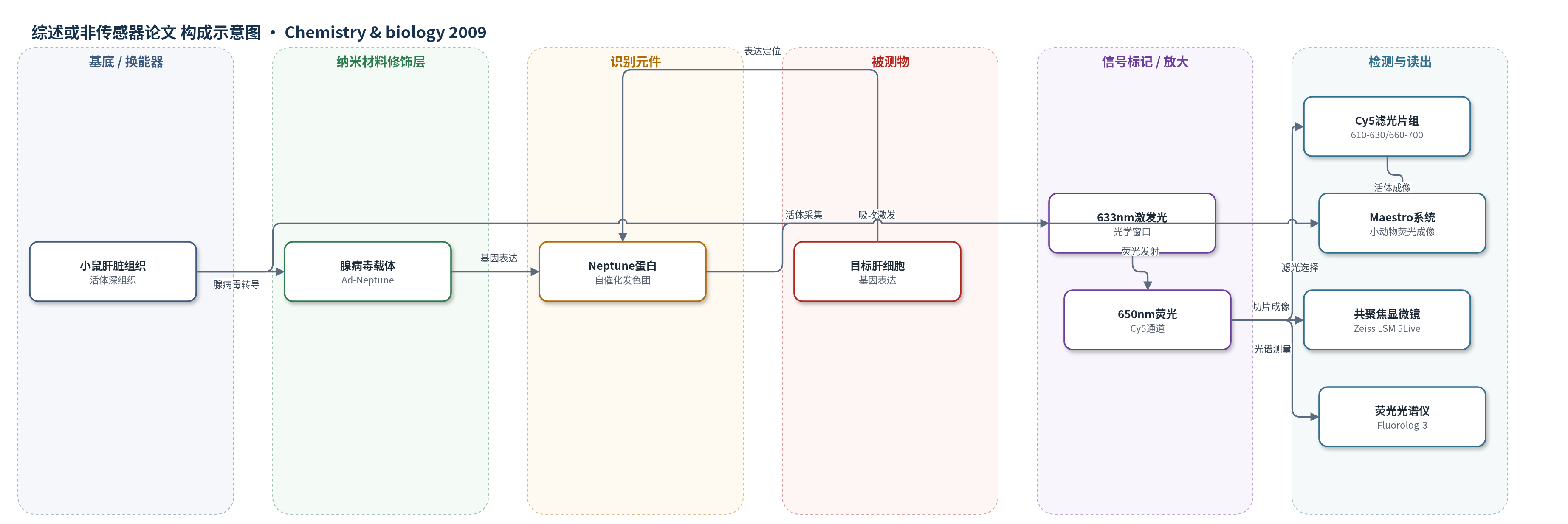
点击图片查看大图 · 依据论文自动绘制
传感器类型
综述或非传感器论文
检测对象
特定细胞群体/基因表达(cell population/gene expression,Neptune 报告基因),样品基质:活体小鼠肝脏、肝组织切片、HeLa 细胞
检测原理
腺病毒载体将 Neptune 基因导入小鼠肝细胞,Neptune 在细胞内自催化形成共轭发色团。当 610–633 nm 光学窗口光照射时,发色团吸收光子,电子从最高占据分子轨道跃迁至最低未占轨道,随后发射约 650 nm 远红荧光。目标细胞中 Neptune 表达量越高,荧光强度越强。结构上,M41G 使水分子进入空腔并与发色团酰亚胺氧形成氢键,稳定激发态并降低 HOMO-LUMO 能隙;Arg197 重排和 Cys158 使发色团环共平面性增强,进一步红移激发与发射。该机制无需外源辅因子,适合活体深组织成像。
检测灵敏度
原文未报告 LOD、线性范围、灵敏度斜率或相关系数。
效应效果
Neptune 激发峰 600 nm、发射峰 650 nm,消光系数 72,000 M^-1 cm^-1,量子产率 0.18,633 nm 激发亮度 1.7(相对单位),光漂白半衰期 185 s,pKa 5.8,成熟半时间 35 min。mNeptune 消光系数 67,000 M^-1 cm^-1,量子产率 0.20,亮度 1.8,光漂白 160 s,解离常数 2.1 mM(Neptune 0.5 mM)。活鼠肝脏 Cy5 通道对比未感染组织自荧光 44 倍;与 IFP1.1 正交成像,交叉信号可线性解混。mNeptune-actin/tubulin 在 HeLa 细胞正确定位,4 个月发色团水解 <7%。可用于活体追踪细胞群体、肿瘤转移和干细胞治疗。
传感器的构成
- 成像探针:Neptune/mNeptune 远红荧光蛋白,自催化形成发色团,作为基因表达荧光标记
- 表达载体:腺病毒载体 Ad-Neptune/Ad-mKate,携带 CMV 启动子和 IRES-GFP,用于小鼠肝细胞转导
- 激发光源:610–630 nm 或 633 nm 激光/氙灯,位于组织光学窗口
- 滤光片组:Cy5 滤光片(激发 610–630 nm,发射 660–700 nm)和 Cy5.5 滤光片(激发 625–675 nm,发射 700–730 nm)
- 读出设备:Maestro 1 小动物荧光成像系统、Zeiss LSM 5Live 共聚焦显微镜、Fluorolog-3 荧光光谱仪
中文摘要
荧光蛋白已成为生物医学研究中重要的蛋白标签、基因表达报告分子、生物传感器组分和细胞谱系追踪工具。然而,由于血红蛋白对 600 nm 以下激发光的强吸收,哺乳动物深组织活体成像受到限制,因此需要能在 600 nm 以上“光学窗口”内有效激发的荧光蛋白。本文通过定点突变和随机突变定向进化,从 Discosoma 和 Entacmaea 家族红色荧光蛋白出发,获得激发峰位于 600 nm 或以上的远红荧光蛋白。其中亮度最高的 Neptune 激发峰为 600 nm、发射峰为 650 nm,在活鼠深组织成像中表现良好。Neptune 的晶体结构揭示了新的红移机制:M41G 突变引入的水分子与发色团酰亚胺区域形成氢键,同时发色团共平面性增强,使激发和发射光谱均发生红移。
英文摘要
Fluorescent proteins have become valuable tools for biomedical research as protein tags, reporters of gene expression, biosensor components, and cell lineage tracers. However, applications of fluorescent proteins for deep tissue imaging in whole mammals have been constrained by the opacity of tissues to excitation light below 600 nm, because of absorbance by hemoglobin. Fluorescent proteins that excite efficiently in the "optical window" above 600 nm are therefore highly desirable. We report here the evolution of far-red fluorescent proteins with peak excitation at 600 nm or above. The brightest one of these, Neptune, performs well in imaging deep tissues in living mice. The crystal structure of Neptune reveals a novel mechanism for red-shifting involving the acquisition of a new hydrogen bond with the acylimine region of the chromophore.