组成图示
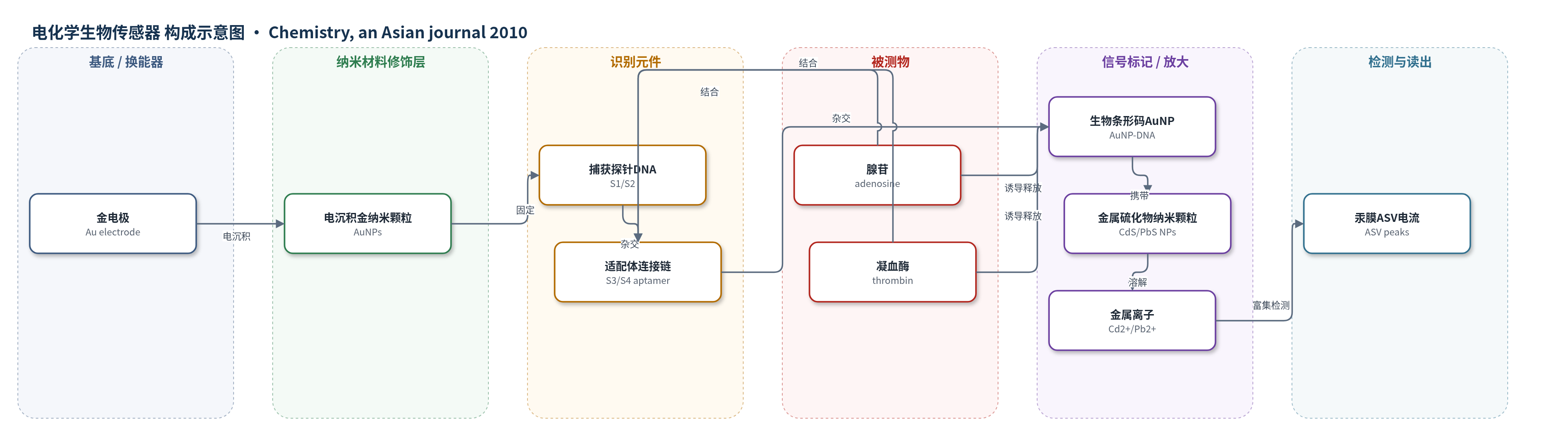
点击图片查看大图 · 依据论文自动绘制
传感器类型
电化学生物传感器
检测对象
腺苷(adenosine)、凝血酶(thrombin);样品基质:人血清/血浆(serum/plasma)
检测原理
金电极表面固定两种巯基捕获探针,连接DNA(S3/S4)含腺苷或凝血酶适配体,并与AuNP上的报告DNA预杂交,形成三明治界面。加入腺苷或凝血酶后,适配体与目标特异性结合并折叠,使连接DNA与报告DNA间的杂交解离,携带CdS或PbS纳米颗粒的生物条形码AuNP释放到溶液中。随后用HNO3溶解金属硫化物纳米颗粒,得到Cd2+或Pb2+,在汞膜电极上经ASV富集和阳极剥离,峰电流随目标浓度升高而增大。该策略利用AuNP生物条形码多探针放大和ASV金属离子预富集,实现信号开启型双目标检测。
检测灵敏度
LOD: 6.6×10^-12 m (adenosine), 1.0×10^-12 m (thrombin);线性范围: 2.0×10^-11–2.0×10^-9 m (adenosine), 2.0×10^-12–2.0×10^-10 m (thrombin);斜率: 0.3215 (adenosine, x为10^-11 m), 0.3070 (thrombin, x为10^-12 m);R^2: 0.9999 (adenosine), 0.9998 (thrombin)
效应效果
传感器选择性良好:单独腺苷仅产生Cd峰,单独凝血酶仅产生Pb峰;胞嘧啶核苷和尿苷(2.0×10^-10 m)、BSA和溶菌酶(2.0×10^-11 m)无明显干扰。对6.0×10^-11 m腺苷和6.0×10^-12 m凝血酶重复8次,Cd和Pb峰相对偏差分别为8.4%和6.3%。加标回收率为腺苷82.2–115.4%、凝血酶85.3–118.6%。六份患者血浆中腺苷为55.2–90.8 nm,四份检出低纳摩尔凝血酶(2.2、3.8、4.7、2.5 nm)。其检出限低于此前1.8×10^-10 m腺苷方法及文献单/双目标适配体传感器,作者认为可用于生物流体中小分子与蛋白同时检测。
传感器的构成
- 基底电极:金电极(Au electrode),作为传感表面与换能器
- 纳米修饰层:直接电沉积金纳米颗粒(AuNPs),增大电极有效表面积
- 识别捕获层:巯基化捕获探针DNA(S1、S2),通过硫-金亲和固定并分别识别腺苷/凝血酶适配体连接链
- 封闭规整层:6-巯基-1-己醇(MCH),规整DNA单分子层并封闭非特异位点
- 识别元件层:连接DNA(S3、S4),含腺苷适配体或凝血酶适配体及扩展序列,介导目标结合与界面释放
- 信号标记层:生物条形码金纳米颗粒(AuNPs)负载报告DNA(S5、S6)和信号DNA(S7),并偶联CdS/PbS纳米颗粒
- 信号读出层:硝酸(HNO3)溶解金属硫化物纳米颗粒,阳极剥离伏安法(ASV)测定Cd2+/Pb2+
中文摘要
本文报道了一种基于适配体构象切换的信号开启型电化学传感策略,用于同时检测腺苷和凝血酶。以金电极为传感表面,修饰两种巯基化捕获探针,分别与含有腺苷适配体或凝血酶适配体的连接DNA互补。连接DNA预先与负载于金纳米颗粒(AuNPs)上的报告DNA杂交,AuNPs同时携带与连接DNA互补的报告DNA和标记不同金属硫化物纳米颗粒的信号DNA,从而构建腺苷与凝血酶的三明治型传感界面。加入腺苷和凝血酶后,适配体部分与目标结合并折叠形成复合物,导致生物条形码AuNPs释放到溶液中。用阳极剥离伏安法(ASV)测定金属硫化物纳米颗粒,腺苷和凝血酶浓度分别与相应金属离子信号成正比。借助生物条形码AuNPs和ASV金属离子预富集的双重放大,腺苷和凝血酶检出限分别低至6.6×10^-12 m和1.0×10^-12 m,传感器在生物样品中表现出优异的选择性和检测能力。
英文摘要
In the present work, a signal-on electrochemical sensing strategy for the simultaneous detection of adenosine and thrombin is developed based on switching structures of aptamers. An Au electrode as the sensing surface is modified with two kinds of thiolated capture probes complementary to the linker DNA that contains either an adenosine aptamer or thrombin aptamer. The capture probes hybridize with their corresponding linker DNA, which has prehybridized with the reporter DNA loaded onto the gold nanoparticles (AuNPs). The AuNP contained two kinds of bio-barcode DNA: one is complementary to the linker DNA (reporter), whereas the other is not (signal) and is tagged with different metal sulfide nanoparticles. Thus a "sandwich-type" sensing interface is fabricated for adenosine and thrombin. With the introduction of adenosine and thrombin, the aptamer parts bind with their targets and fold to form the complex structures. As a result, the bio-barcoded AuNPs are released into solution. The metal sulfide nanoparticles are measured by anodic stripping voltammetry (ASV), and the concentrations of adenosine and thrombin are proportional to the signal of either metal ion. With the dual amplification of the bio-barcoded AuNP and the preconcentration of metal ions through ASV technology, detection limits as low as 6.6 x 10(-12) M for adenosine and 1.0 x 10(-12) M for thrombin are achieved. The sensor exhibits excellent selectivity and detectability in biological samples.