组成图示
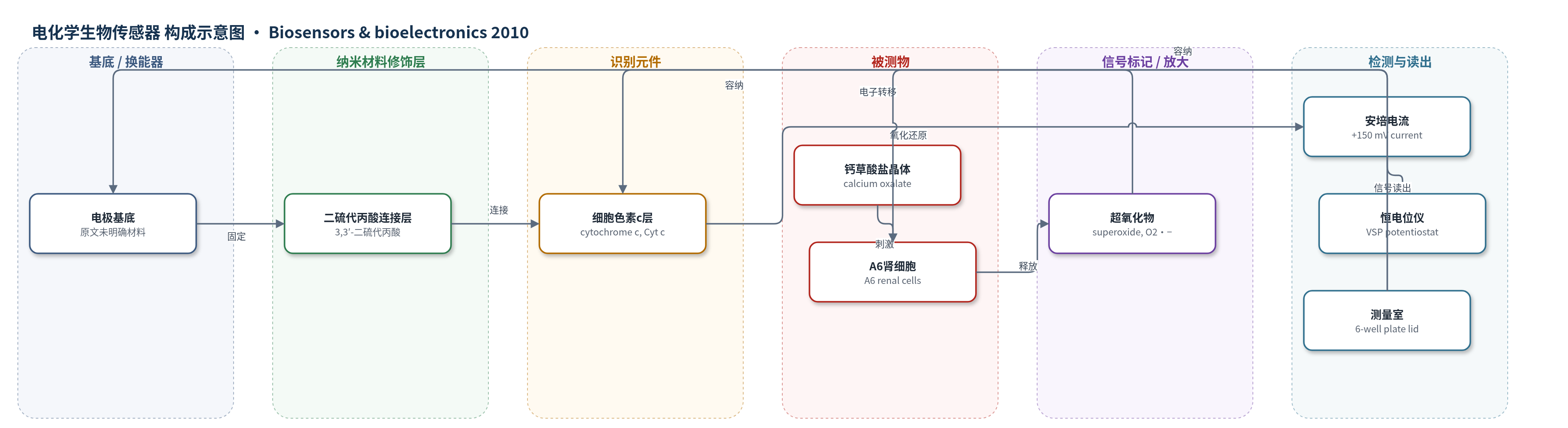
点击图片查看大图 · 依据论文自动绘制
传感器类型
电化学生物传感器
检测对象
超氧化物(superoxide, O2•−);样品基质:A6 肾细胞胞外空间(Ringer buffer 培养液)
检测原理
该传感器以表面固定细胞色素 c 作为识别/电子转导元件。钙草酸盐晶体刺激 A6 肾细胞后,细胞通过 NAD(P)H 氧化酶等途径向胞外空间释放超氧化物 O2•−。O2•− 扩散至电极表面,将细胞色素 c 还原;在 +150 mV 恒电位下,被还原的细胞色素 c 在电极上重新氧化,产生与 O2•− 浓度成正比的安培电流。传感器通过 3,3′-二硫代丙酸连接层将细胞色素 c 固定于电极,保持电子转导并降低 H2O2、抗坏血酸、尿酸等干扰。校准采用黄嘌呤氧化酶/次黄嘌呤体系酶促生成 O2•−,使电流可换算为绝对浓度。无额外酶催化放大,信号直接反映胞外 O2•− 瞬时浓度。
检测灵敏度
灵敏度: 18.3 ± 2.7 × 10^2 A m−2 M−1;线性范围: ≤1 μM superoxide
效应效果
该传感器对 H2O2、抗坏血酸和尿酸等常见干扰物具有较低干扰,加入超氧化物歧化酶(SOD)后校准电流降低 85–100%,细胞实验中 SOD 使电流由 2.6 nA 降至 0.3 nA,表明超过 90% 信号来自超氧化物。传感器达到稳态电流的时间为 27.1 ± 11.4 s,可实现优于 1 min 的时间分辨率;表面固定细胞色素 c 浓度为 11.7 ± 2.9 pmol cm−2,灵敏度为 18.3 ± 2.7 × 10^2 A m−2 M−1,线性可达 1 μM。作者强调该系统可连续监测短寿命活性氧,避免终点法无法反映瞬时氧化爆发的问题,并可结合 NADPH 氧化酶抑制剂等用于研究肾结石相关细胞损伤。
传感器的构成
- 换能器电极:原文未明确具体材料(仅称 electrode),作为电子转导基底并输出安培电流
- 连接/间隔层:3,3′-dithiodipropionic acid,作为电极与细胞色素 c 之间的连接分子,提供表面官能团并维持电子转导
- 识别/电子转导元件:cytochrome c (Cyt c),表面固定单层,接受超氧化物电子并发生氧化还原
- 被测物:superoxide (O2•−),由 A6 肾细胞在钙草酸盐刺激下释放至胞外空间
- 测量室:modified 6-well plate lid 与 electrode system,用于将传感器定位在细胞附近并实时监测
- 工作介质:Ringer buffer (pH 7.4),提供离子环境并维持细胞与传感器工作
中文摘要
钙草酸盐是肾结石的主要成分之一,已被证实通过氧化应激对肾细胞产生有害作用。然而,这种氧化应激的亚细胞来源以及是否延伸至胞外空间仍存在争议。为此,作者构建了一种电化学超氧化物生物传感器,并将其置于 A6 肾细胞上方,用于实时监测加入钙草酸盐晶体后细胞胞外空间的氧化爆发。结果表明,A6 细胞在接触钙草酸盐晶体后数分钟内即向胞外空间分泌超氧化物,释放量在约 20 min 达到峰值,并在约 3 h 后从胞外空间清除。超氧化物分泌受超氧化物歧化酶存在与否、细胞培养龄期、钙草酸盐晶体用量及温度等因素影响。此外,佛波醇 12-肉豆蔻酸酯 13-乙酸酯(PMA)可模拟钙草酸盐晶体诱导的超氧化物释放。所开发的传感系统可为生物学家在细胞水平研究肾结石形成机制提供有用工具。
英文摘要
Calcium oxalate is one of the main constituents of kidney stones and has a proved deleterious effect on renal cells that is mediated by oxidative stress. However, the subcellular source of this oxidative stress, and whether it is extending to the extracellular space or not, is still disputed. Therefore, an electrochemical superoxide biosensor was constructed, positioned above A6 renal cells, and used to measure in real-time the extracellular oxidative burst following addition of calcium oxalate crystals. It was observed that A6 cells do secrete superoxide into their extracellular space in few minutes after encountering calcium oxalate crystals. The amount of released superoxide peaks at about 20 min. Superoxide is cleared away from the extracellular space after approximately 3h. Superoxide secretion depends on the presence of superoxide-scavenging enzyme superoxide dismutase, the age of the cells, the amount of calcium oxalate crystals, and the temperature. Moreover, the effect of calcium oxalate crystals was mimicked by phorbol 12-myristate 13-acetate. The developed sensing system can be a useful tool for biologists investigating nephrolithiasis at cellular level.