组成图示
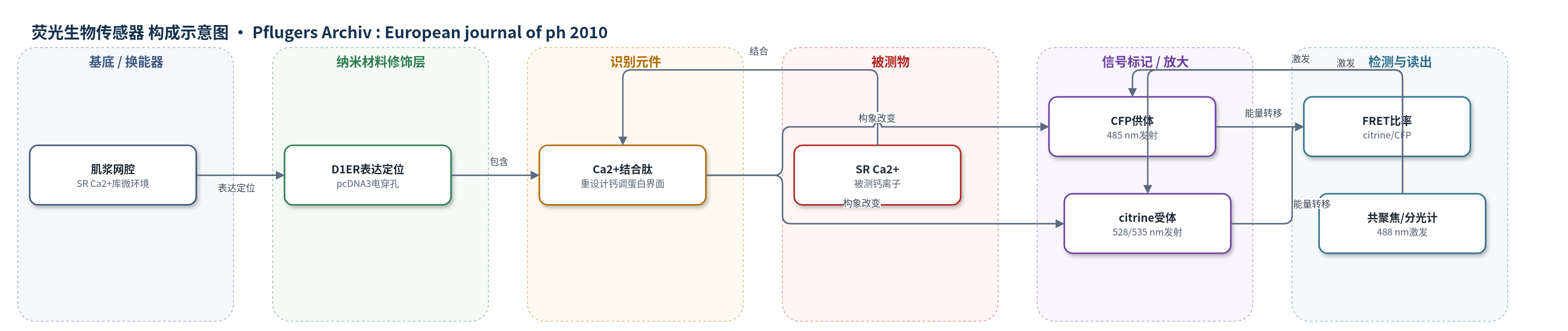
点击图片查看大图 · 依据论文自动绘制
传感器类型
荧光生物传感器
检测对象
肌浆网钙离子(SR Ca2+);样品基质为成年小鼠屈指短肌(FDB)骨骼肌纤维的肌浆网腔
检测原理
D1ER 为靶向肌浆网(SR)的 cameleon FRET 钙传感器,由 CFP 供体、citrine 受体及重设计的钙调蛋白结合肽组成。SR 腔内 Ca2+ 与结合肽结合后改变 CFP 与 citrine 的相对距离/取向,使 FRET 效率升高,CFP 发射降低、citrine 发射升高,citrine/CFP 比率升高;当电刺激或 4-CmC 激活 RyR1 引起 SR Ca2+ 释放时,Ca2+ 解离,FRET 降低,citrine/CFP 比率下降。通过 488 nm 激发并双通道记录 citrine 与 CFP 发射,比率化读出可部分抵消探针浓度和光漂白影响,从而反映 SR Ca2+ 相对变化与耗竭程度。
检测灵敏度
r2=0.6;Pearson correlation=0.776, p=0.0025
效应效果
D1ER 与 di-8-ANEPPS 标记的 T 管/三联体共定位,ryanodine 抑制 FRET 下降,提示信号来自 RyR1 介导的 SR 钙释放。基线 citrine/CFP 为 1.62±0.10(0.39–7.40,n=100),48 h 稳定;仅 17/32(53%)纤维响应电刺激。100 ms 去极化至 20 mV 平均降低 6.45±0.8%(2.6–11.7%),4-CmC 平均降低 20.6±3.6%(6.6–57.4%)。电压依赖符合 Boltzmann 函数,与 Fura-2FF 胞质钙信号相近。相比 Fluo-5 N、SEER,D1ER 可原位比率化监测 SR 钙,但校准和动力学有限。
传感器的构成
- 表达载体:pcDNA3 质粒携带 D1ER cameleon 基因,经电穿孔进入 FDB 肌纤维并驱动 D1ER 表达
- 传感微环境:肌浆网(SR)腔/终末池,提供高 Ca2+ 微环境并作为 D1ER 定位位点
- 识别元件:D1ER 中重设计的钙调蛋白结合肽/钙调蛋白界面,结合 Ca2+ 并改变构象
- 荧光供体:CFP(cyan fluorescent protein),作为 FRET 供体,发射约 485 nm
- 荧光受体:citrine(黄色荧光蛋白),作为 FRET 受体,发射约 528/535 nm
- 读出装置:共聚焦显微镜/分光荧光计,488 nm 激发并以双光电倍增管记录 citrine/CFP 比率
中文摘要
内质网/肌浆网(ER/SR)在多种细胞胞质信号中起关键作用,对骨骼肌纤维尤为重要,该细胞器是收缩等基本功能所需的主要钙库。本研究通过体内电穿孔在小鼠屈指短肌(FDB)中表达 cameleon 生物传感器 D1ER,直接评估电刺激和药理学刺激下 SR Ca2+ 耗竭。主要结论:(1)D1ER 在 FDB 纤维 SR 中表达,di-8-(氨基萘乙烯基吡啶) 染色和 SR Ca2+ 释放后 FRET 信号降低均支持该定位;(2)4-氯间甲酚(4-CmC)或电刺激诱发的 D1ER citrine/CFP 比率幅度与基线 citrine/CFP 比率直接成正比,表明完整肌纤维中 SR Ca2+ 调节 RyR1 介导的 SR Ca2+ 释放;(3)以 D1ER citrine/CFP 信号测得的 SR Ca2+ 释放具有电压依赖性,符合 Boltzmann 函数;(4)4-CmC 和持续肌膜去极化引起的平均 SR Ca2+ 耗竭分别为 20% 和 6.4%。结果表明,在生理条件下显著耗竭 SR Ca2+ 含量较难。
英文摘要
The endoplasmic/sarcoplasmic reticulum (ER/SR) plays a crucial role in cytoplasmic signalling in a variety of cells. It is particularly relevant to skeletal muscle fibres, where this organelle constitutes the main Ca2+ store for essential functions, such as contraction. In this work, we expressed the cameleon biosensor D1ER by in vivo electroporation in the mouse flexor digitorum brevis (FDB) muscle to directly assess SR Ca2+ depletion in response to electrical and pharmacological stimulation. The main conclusions are: (1) D1ER is expressed in the SR of FDB fibres according to both di-8-(amino naphthyl ethenyl pyridinium) staining experiments and reductions in the Förster resonance energy transfer signal consequent to SR Ca2+ release; (2) the amplitude of D1ER citrine/cyan fluorescent protein (CFP) ratio evoked by either 4-chloro-m-cresol (4-CmC) or electrical stimulation is directly proportional to the basal citrine/CFP ratio, which indicates that SR Ca2+ modulates ryanodine-receptor-isoform-1-mediated SR Ca2+ release in the intact muscle fibre; (3) SR Ca2+ release, measured as D1ER citrine/CFP signal, is voltage-dependent and follows a Boltzmann function; and (4) average SR Ca2+ depletion is 20% in response to 4-CmC and 6.4% in response to prolonged sarcolemmal depolarization. These results indicate that significantly depleting SR Ca2+ content under physiological conditions is difficult.