组成图示
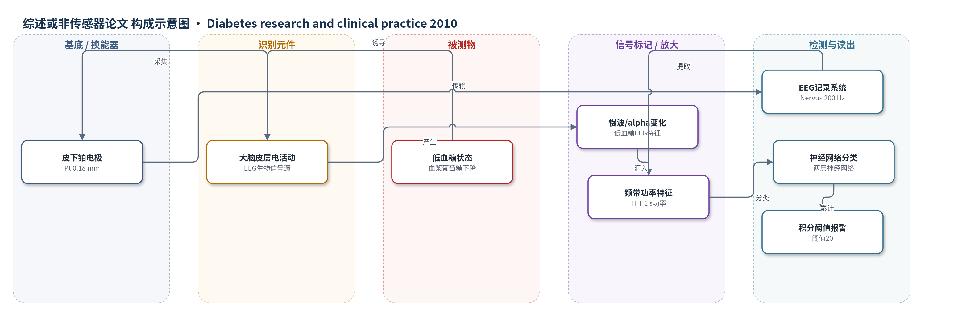
点击图片查看大图 · 依据论文自动绘制
传感器类型
综述或非传感器论文
检测对象
低血糖(hypoglycemia,血浆葡萄糖下降,plasma glucose);样品基质:1型糖尿病患者体内/血浆(胰岛素诱导低血糖)
检测原理
胰岛素诱导血浆葡萄糖下降,脑神经元代谢受抑,皮层EEG出现alpha活动减少、慢波(theta/delta)增加。皮下铂电极将皮层电位转换为电信号,EEG系统200 Hz采样。算法对1 s片段做FFT,提取alpha、beta、gamma、theta功率及噪声指标;两层神经网络基于贝叶斯学习识别低血糖一致片段。随后用10 min线性衰减核积分事件,累计曲线超过预定义阈值20即报警。低血糖越重或持续越久,慢波片段越多,积分值越高,从而在严重认知障碍前给出预警。
检测灵敏度
相关系数: r^2 = 0.82, p = 0.046(Spearman相关)
效应效果
在15名1型糖尿病患者中,自动算法100%检测到胰岛素诱导低血糖相关EEG变化。EEG积分曲线超过阈值时血浆葡萄糖为2.0–3.4 mmol/L,平均早于停止胰岛素输注29±28 min(范围3–113 min);12/15早于认知测试定义的严重低血糖,14/15预警间隔≥9 min。葡萄糖输注后积分曲线平均10.3 min降至阈值以下(14/15)。重复研究日个体内血糖阈值CV为8%,组间CV为14%,两天阈值血糖相关r^2=0.82(p=0.046)。24 h以上正常血糖EEG仅1次疑似假阳性。预警时间与年龄、病程、HbA1c及降糖速率无相关,glucagon应答者与非应答者无差异。作者认为皮下电极+自动算法可发展为日常携带的低血糖报警装置。
传感器的构成
- 换能器电极:4枚皮下铂电极(Pt,直径0.18 mm,Ad-Tech),采集皮层电活动
- 信号采集系统:Nervus EEG记录系统,200 Hz采样,传输电极信号
- 生物识别源:大脑皮层神经元电活动,低血糖时出现慢波增加和alpha活动减少
- 特征提取层:1 s EEG片段FFT功率估计,提取alpha、beta、gamma、theta频带功率及噪声指标
- 分类模型:两层神经网络(双曲正切函数),基于贝叶斯学习训练,识别低血糖一致EEG片段
- 积分阈值层:10 min线性衰减积分核,累计事件曲线,预定义阈值20触发低血糖报警
中文摘要
目的:低血糖无感知是1型糖尿病患者严重低血糖的常见危险因素。本研究检验以下假设:低血糖期间脑电图(EEG)的特异性变化可由皮下电极记录,并通过通用数学算法处理,且低血糖相关EEG变化出现在严重低血糖发生之前。方法:15名1型糖尿病患者接受胰岛素诱导低血糖并记录EEG,同时以重复认知测试评估认知功能。当血浆葡萄糖降至1.8 mmol/L或受试者出现明显认知功能障碍时停止胰岛素输注。EEG采用预定义低血糖阈值的自动数学算法分析。结果:数学算法在所有受试者中均检测到低血糖相关EEG变化。EEG变化超过阈值时血浆葡萄糖为2.0–3.4 mmol/L,发生在停止胰岛素输注前29±28 min(范围3–113 min)。结论:自动数学算法可检测所有胰岛素诱导低血糖受试者的低血糖相关EEG变化;15人中12人的EEG变化早于认知测试评估的严重低血糖。
英文摘要
AIMS: Hypoglycemia unawareness is a common condition associated with increased risk of severe hypoglycemia. We test the hypothesis that specific changes in the electroencephalogram (EEG) during hypoglycemia can be recorded by subcutaneous electrodes and processed by a general mathematical algorithm, and that hypoglycemia associated EEG changes appear before the development of severe hypoglycemia.
METHODS: Fifteen patients with type 1 diabetes were exposed to insulin-induced hypoglycemia and EEG was recorded. The cognitive function was evaluated by repeated cognitive testing. Insulin infusion was terminated when plasma glucose reached 1.8mmol/l or when the subjects showed obvious signs of cognitive dysfunction. EEG was analyzed by an automated mathematical algorithm with a predefined threshold of hypoglycemia.
RESULTS: Hypoglycemia associated EEG changes were detected by the mathematical algorithm in all subjects. Plasma glucose at the time of EEG changes above the threshold value ranged from 2.0 to 3.4mmol/l and occurred 29+/-28min (range 3-113min) before termination of insulin infusion.
CONCLUSIONS: Hypoglycemia associated EEG changes could be detected by an automated mathematical algorithm in all subjects exposed to insulin-induced hypoglycemia. In 12 of 15 patients, EEG changes occurred before severe hypoglycemia as evaluated by the cognitive testing.