组成图示
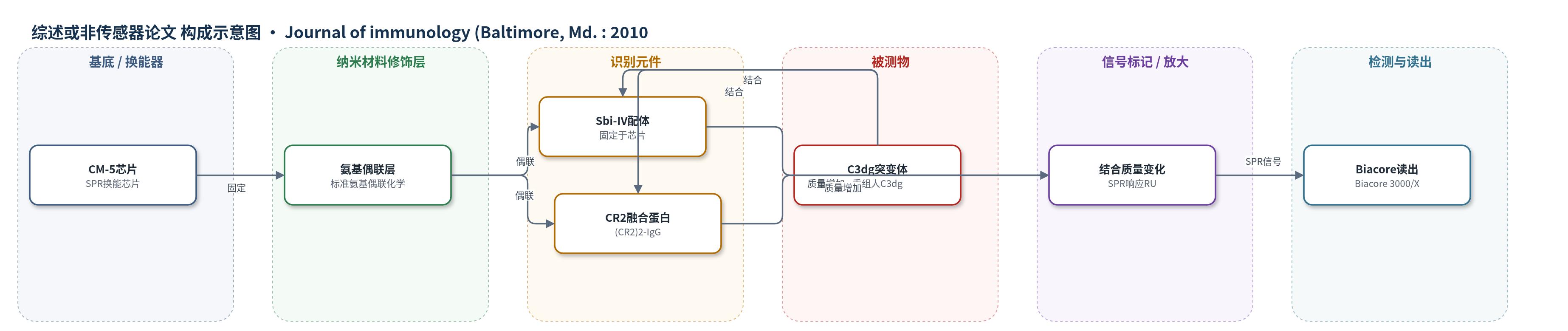
点击图片查看大图 · 依据论文自动绘制
传感器类型
综述或非传感器论文
检测对象
重组人 C3dg(C3dg)及其点突变体;样品基质:HBS-E 缓冲液(含 0.005% P20)中的重组蛋白溶液;另以 Sbi-IV 作为竞争共分析物。
检测原理
该研究将 Sbi-IV 或 (CR2)2-IgG 通过氨基偶联固定于 CM-5 SPR 芯片,形成识别层。向流动池注入重组 C3dg 或其突变体后,C3dg 与固定配体结合,使芯片表面质量/折射率发生变化,Biacore 以响应单位(RU)实时读出。稳态 RU 随 C3dg 浓度升高而增加,可用单位点 Langmuir 等温线拟合得到结合常数。若 C3d 凹面酸性口袋关键残基被突变,结合减弱,RU 平台下降;侧沟酸性残基或 N170 突变则基本不影响结合。Sbi-IV 与 CR2 结合位点重叠,因此 Sbi-IV 作为共分析物可竞争抑制 C3dg-CR2 结合,信号随 Sbi-IV 过量而降低。
检测灵敏度
原文未报告 LOD、线性范围、灵敏度斜率或相关系数。
效应效果
SPR 在 25°C、HBS-E 含 0.005% P20 条件下进行,Sbi-IV 固定约 1100 RU,(CR2)2-IgG 固定 500–4800 RU;C3dg 浓度 0.1 mg/ml(原文标 2.5 mM)。野生型 C3dg 与固定 (CR2)2-IgG 的 KD 为 9.4 mM(原文单位)。D36A、R49M 使 Sbi-IV 结合几乎完全丧失;D163A、I164A 使 CR2 结合降至野生型约 5%、8%。侧沟 E117A/D122A/D128A/D147A 及 N170A 无明显影响。0.2 M 锌醋酸盐基本消除 CR2-C3dg 结合,提示共晶条件可能非生理。未报告 RSD、回收率或临床验证,但为 CR2/C3d 界面争议提供独立 SPR 证据。
传感器的构成
- 基底/换能器:CM-5 生物传感器芯片(SPR 换能芯片),用于固定配体并产生表面等离子共振信号
- 配体固定层:标准氨基偶联化学形成的共价偶联层,用于将 Sbi-IV 或 (CR2)2-IgG 固定于芯片
- 识别元件:Sbi-IV(金黄色葡萄球菌 Sbi 第 IV 结构域)或 (CR2)2-IgG 融合蛋白(含 CR2 SCR1-2),作为结合 C3dg 的配体
- 分析物:重组人 C3dg 及其点突变体,以 0.1 mg/ml(原文标为 2.5 mM)注入流动池
- 缓冲/封闭:HBS-E(10 mM HEPES、0.15 M NaCl、3 mM EDTA,pH 7.2)含 0.005% P20 表面活性剂,维持结合条件并减少非特异吸附
- 信号读出:Biacore 3000 或 Biacore-X SPR 仪器,以响应单位(RU)监测结合稳态与动力学
中文摘要
作者近期报道金黄色葡萄球菌免疫逃逸分子Sbi与补体C3的相互作用,主要由C3d(g)结合Sbi第IV结构域(Sbi-IV)介导。基于这些研究,作者通过定点突变分析C3d与Sbi-IV的结合界面,并重新审视C3d上补体受体2(CR2)结合位点的争议。此前发现Sbi III-IV片段结合C3dg会抑制C3dg与CR2结合;而C3d与Efb-C(Sbi-IV的结构/功能同源物)共晶结构显示Efb-C结合于C3d凹面区域,该区域曾被作者突变数据提示参与CR2结合,但未在CR2(SCR1-2):C3d共晶结构中证实。本研究用表面等离子共振(SPR)分析一系列C3dg突变体与生物传感器固定的Sbi-IV或CR2(SCR1-2)的结合。结果显示,C3d凹面酸性口袋突变显著影响与Sbi-IV和CR2的结合,但两者最关键残基存在差异;而共晶结构提示的侧沟突变不引起结合缺陷。Sbi-IV结果提示其结合模式与Efb-C:C3d复合物高度相似;CR2结果证实早期定位研究,并进一步质疑C3d:CR2共晶结构的生理相关性。
英文摘要
We recently characterized an interaction between the Staphylococcus aureus immune evasion molecule Staphylococcus aureus binder of Ig (Sbi) and complement C3, an interaction mediated primarily through the binding of C3d(g) to Sbi domain IV. Events related to these studies prompted us to investigate via mutagenesis the binding interface of C3d for Sbi domain IV (Sbi-IV), as well as to revisit the controversial issue of the complement receptor 2 (CR2) binding site of C3d. Specifically, we had shown that Sbi domains III and IV fragment binding to C3dg inhibited the latter's binding to CR2. Moreover, a published cocrystal structure of C3d bound to complement inhibitory C-terminal domain of extracellular fibrinogen-binding protein (Efb-C), a structural and functional homolog of Sbi-IV, showed Efb-C binding to a region on the concave face of C3d previously implicated in CR2 binding by our mutagenesis data but not confirmed in the CR2(short consensus repeat [SCR]1-2):C3d cocrystal structure. We have now analyzed by surface plasmon resonance the binding of a series of variant C3dg molecules to biosensor-bound Sbi-IV or CR2(SCR1-2). We found that mutations to the concave face acidic pocket of C3d significantly affected binding to both Sbi-IV and CR2, although there was divergence in which residues were most important in each case. By contrast, no binding defects were seen for mutations made to the sideface of C3d implicated from the cocrystal structure to be involved in binding CR2(SCR1-2). The results with Sbi-IV suggest a mode of binding highly similar to that visualized in the Efb-C:C3d complex. The results with CR2 confirm our earlier mapping studies and cast even further doubt on the physiologic relevance of the complex visualized in the C3d:CR2 cocrystal.