组成图示
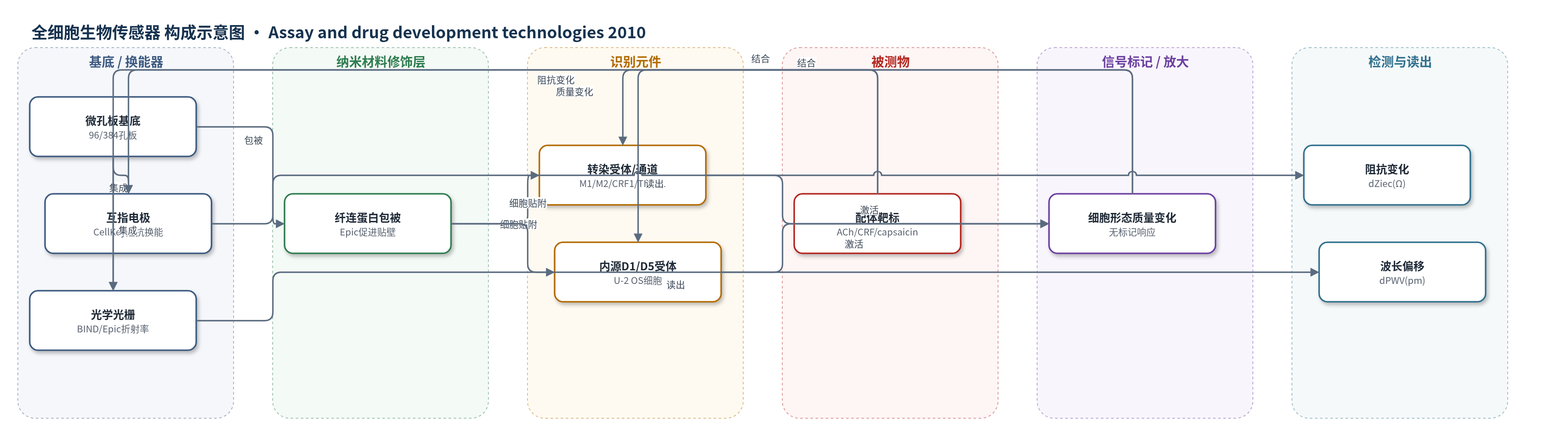
点击图片查看大图 · 依据论文自动绘制
传感器类型
全细胞生物传感器
检测对象
乙酰胆碱(ACh)、卡巴胆碱(carbachol)、促肾上腺皮质激素释放因子(CRF)、sauvagine、辣椒素(capsaicin)、二氢雷替定(DHX)、forskolin、α-MSH、AgRP 等 GPCR/TRPV1 配体;样品基质为 CHO-K1 或 U-2 OS 活细胞培养孔板(HBSS 缓冲液)。
检测原理
细胞贴附于微孔板换能表面后,配体与细胞表面 GPCR 或 TRPV1 结合,激活 Gi、Gq、Gs 信号或离子通道,引起细胞骨架重排、形态、黏附和近表面质量变化。CellKey 通过互指电极测量细胞层阻抗,配体浓度升高使 dZiec 改变;Gi 通常产生正响应,Gs 产生负响应,Gq 先短暂下降后上升。BIND 和 Epic 通过光学光栅检测近表面折射率/质量变化,反射光波长偏移 dPWV 随配体浓度变化。系统无需标记配体或受体,利用高灵敏度检测 nm 级细胞微运动或约 300 Da 质量变化,峰响应用于 4 参数逻辑拟合。
检测灵敏度
—
效应效果
三种仪器在激动剂和拮抗剂模式下均稳健,Z′ 值为 0.5–0.8(CRF1:0.6/0.8/0.5;M1:0.8/0.8/0.5;M2:0.7/0.7/0.6),重现性以较小 SEM 和可比方差体现。各平台 pEC50/pIC50 可比,如 DHX 为 8.8/9.1,forskolin 为 6.9/6.8,AgRP 为 7.3/7.6;CRF1 低效相 pEC50 为 6.8/7.3/7.8。选择性由拮抗剂抑制、未转染细胞无响应及 cytochalasin D 降低响应支持。CellKey 可定性区分 Gi、Gs、Gq 并揭示 CRF1 双重信号;光学平台可检测反向激动。作者认为其适合活细胞药物发现。
传感器的构成
- 基底/换能器:CellKey 96 孔微孔板内嵌互指电极(interdigitated electrodes),施加小电流测量细胞层阻抗;BIND/Epic 384 孔微孔板表面集成光学光栅(optical grating),检测近表面折射率/质量变化。
- 表面修饰:Epic 使用纤连蛋白(fibronectin)包被 384 孔板促进细胞贴壁;BIND 使用 CA2 孔板;CellKey 使用专用 96 孔板;原文未报告纳米材料修饰。
- 识别元件:CHO-K1 稳定转染人 GPCR/离子通道(M1、M2、CRF1、TRPV1、MC4R)或 U-2 OS 内源 D1/D5 受体,作为细胞表面识别元件。
- 靶标:乙酰胆碱(ACh)、卡巴胆碱(carbachol)、CRF、sauvagine、辣椒素(capsaicin)、DHX、forskolin、MSH、AgRP 等配体,加入细胞培养孔。
- 信号标记物:无外源标记物,依赖受体/通道激活后细胞骨架、形态、黏附和近表面质量变化产生无标记信号。
- 读出:CellKey 输出阻抗变化 dZiec(Ω);BIND/Epic 输出反射光波长偏移 dPWV(pm),用于浓度-响应曲线。
中文摘要
无标记技术在药物发现中日益重要。现有两类无标记仪器可用于研究配体对细胞靶标的影响:电学阻抗型和光学型。两类生物传感器对细胞形态和结构变化均高度敏感,但比较同一 GPCR 响应时是否结果相似尚不清楚;不同 G 蛋白激活引起的细胞形态变化不同,仪器可能对 G 蛋白信号具有不同灵敏度。本研究用 1 台阻抗仪器(CellKey)和 2 台光学仪器(BIND、Epic),比较 Gi 偶联(ACh M2)、Gq 偶联(ACh M1)和 Gs 偶联(CRF1)受体。三种仪器在激动剂和拮抗剂模式下均稳健,效力和方差可比。阻抗和光学平台对内源 D1/D5 受体响应及黑皮质素 4 受体反向激动剂 AgRP 均高灵敏。阻抗平台还能定性区分 G 蛋白偶联并揭示 CRF1 双重信号。TRPV1 响应在三台仪器中均可检测。因此,两类平台均提供稳健活细胞无标记检测,适合药物发现,通常给出相似 GPCR 药理学特征。
英文摘要
The diversity and impact of label-free technologies continues to expand in drug discovery. Two classes of label-free instruments, using either an electrical impedance-based or an optical-based biosensor, are now available for investigating the effects of ligands on cellular targets. Studies of GPCR function have been especially prominent with these instruments due to the importance of this target class in drug discovery. Although both classes of biosensors share similar high sensitivity to changes in cell shape and structure, it is unknown whether these biosensors yield similar results when comparing the same GPCR response. Furthermore, since cell morphology changes induced by GPCRs differ depending on which G-protein is activated, there is potential for these instruments to have differential sensitivities to G-protein signaling. Here 1 impedance (CellKey)- and 2 optical-based instruments (BIND and Epic) are compared using Gi-coupled (ACh M2), Gq-coupled (ACh M1), and Gs-coupled (CRF1) receptors. All 3 instruments were robust in agonist and antagonist modes yielding comparable potencies and assay variance. Both the impedance and optical biosensors showed similar high sensitivity for detecting an endogenous D1/D5 receptor response and a melanocortin-4 receptor inverse agonist (agouti-related protein). The impedance-based biosensor was uniquely able to qualitatively distinguish G-protein coupling and reveal dual signaling by CRF1. Finally, responses with a ligand-gated ion channel, TRPV1, were similarly detectable in each instrument. Thus, despite some differences, both impedance- and optical-based platforms offer robust live-cell, label-free assays well suited to drug discovery and typically yield similar pharmacological profiles for GPCR ligands.