组成图示
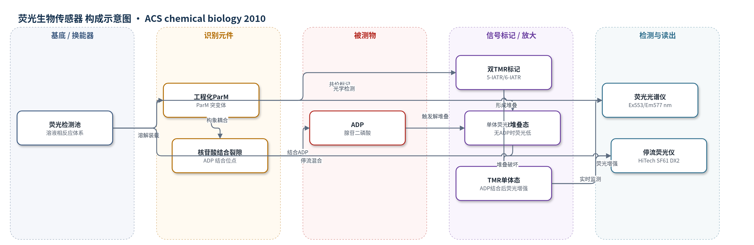
点击图片查看大图 · 依据论文自动绘制
传感器类型
荧光生物传感器
检测对象
ADP(adenosine diphosphate, ADP);样品基质:体外激酶/ATPase 反应缓冲液(含 ATP、底物、Mg2+)
检测原理
该传感器为溶液相蛋白荧光传感器。工程化 ParM 在 Cys63 和 Cys224 位点共价连接两个四甲基罗丹胺(TMR)。无 ADP 时,ParM 处于开放构象,两个 TMR 距离较近并形成堆叠,因激子耦合发生荧光猝灭。ADP 进入 ParM 核苷酸结合裂隙后,诱导两个结构域相对旋转、裂隙闭合,使两个 TMR 的距离和相对取向改变,堆叠态被破坏,TMR 荧光增强约 15 倍。由于传感器对 ADP 的 Kd 约 30 μM,可低于 ADP 浓度使用,信号随 ADP 浓度在 Kd 附近连续变化;对 ATP 的 Kd 大于 3 mM,因此可在毫摩尔 ATP 背景下选择性检测 ADP。荧光在 553 nm 激发、577 nm 发射处读出,停流系统可实时监测结合动力学。
检测灵敏度
动态范围: 亚微摩尔至数十微摩尔 ADP(Kd 附近);线性范围: ≤10 μM ADP(校准近似线性);灵敏度斜率: 6.8 ± 0.2 μM^-1(无 ATP)、5.4 ± 0.1 μM^-1(1 mM ATP)
效应效果
该传感器对 ATP 区分能力 >100 倍,ATP Kd >3 mM,可在毫摩尔 ATP 背景下检测亚微摩尔至数十微摩尔 ADP;1 mM ATP 使校准斜率降低约 20%。盐条件可使 ADP Kd 变化最大约 10 倍,荧光变化 9–15 倍,去除 Mg2+ 后 ADP Kd >5 mM。己糖激酶检测得 KM(ATP)=75±2 μM、Vmax=5.0±0.2 μM min^-1,KM(glucose)=180±9 μM、Vmax=5.4±0.2 μM min^-1,酶活 1.04±0.05 μmol min^-1 unit^-1。AMP 激酶检测得 KM(ATP)=35±3 μM、Vmax=20±2 nM s^-1,KM(SAMS)=36±1 μM、Vmax=19±1 nM s^-1,kcat=9.8±0.6 s^-1。TMR 光稳定性优于香豆素,适合高通量与长光照检测;未报告 RSD/回收率。
传感器的构成
- 基底/换能器:无固体基底,溶液相荧光检测体系(Cary Eclipse 荧光光谱仪/HiTech SF61 DX2 停流系统)
- 识别元件:工程化细菌肌动蛋白同源物 ParM(His6/K33A/D63C/T174A/T175N/D224C/C287A),特异性结合 ADP 并发生构象变化
- 信号标记物:两个四甲基罗丹胺(tetramethylrhodamine, TMR;5-IATR 或 6-IATR)共价连接于 Cys63 与 Cys224,形成可猝灭的堆叠态
- 识别-信号耦合:核苷酸结合裂隙与 TMR 标记位点空间耦合,ADP 结合使两个 TMR 的距离和相对取向改变
- 离子/缓冲环境:30 mM Tris·HCl pH 7.5、25 mM KCl、3 mM MgCl2、5 μM BSA;Mg2+ 为 ADP 结合必需,KCl 调节亲和力
- 读出层:TMR 荧光信号(激发 553 nm、发射 577 nm),由荧光光谱仪或停流荧光仪监测
中文摘要
ADP 荧光检测在基础研究和药物发现中具有重要价值,因为 ADP 是 ATPase 和激酶反应的通用产物。本文报道一种基于四甲基罗丹胺(TMR)标记细菌肌动蛋白同源物 ParM 的新型无试剂荧光生物传感器。该传感器利用 ParM 结合 ADP 时发生的大幅度构象变化,以及 TMR 染料堆叠导致的强荧光猝灭。ParM 在两个邻近位点共价标记两个 TMR,使荧光基团可相互相互作用;ADP 结合改变两个 TMR 的距离和相对取向,从而改变堆叠相互作用并引起荧光强度变化。最终传感器对 ADP 结合产生约 15 倍荧光增强,对 ADP 亲和力较弱(Kd 约 30 μM),因此可相对 ADP 以亚化学计量浓度使用,并在 Kd 附近的亚微摩尔至数十微摩尔范围内报告 ADP 浓度变化。该传感器对 ATP 的区分能力超过 100 倍,可在毫摩尔 ATP 背景下检测 ADP。20 °C 下标记 ParM 结合 ADP 的速率常数为 9.5×10^4 M^-1 s^-1,复合物解离速率为 2.9 s^-1,适合实时测量,并用糖激酶和哺乳动物蛋白激酶验证了其在动力学检测中的性能。
英文摘要
Fluorescence assays for ADP detection are of considerable current interest, both in basic research and in drug discovery, as they provide a generic method for measuring the activity of ATPases and kinases. The development of a novel fluorescent biosensor is described that is based on a tetramethylrhodamine-labeled, bacterial actin homologue, ParM. The design of the biosensor takes advantage of the large conformational change of ParM on ADP binding and the strong quenching of the tetramethylrhodamine fluorescence by stacking of the dye. ParM was labeled with two tetramethylrhodamines in close proximity, whereby the fluorophores are able to interact with each other. ADP binding alters the distance and relative orientation of the tetramethylrhodamines, which leads to a change in this stacking interaction and so in the fluorescence intensity. The final ADP biosensor shows approximately 15-fold fluorescence increase in response to ADP binding. It has relatively weak affinity for ADP (K(d) = 30 microM), enabling it to be used at substoichiometric concentrations relative to ADP, while reporting ADP concentration changes in a wide range around the K(d) value, namely, submicromolar to tens of micromolar. The biosensor strongly discriminates against ATP (>100-fold), allowing ADP detection against a background of millimolar ATP. At 20 degrees C, the labeled ParM binds ADP with a rate constant of 9.5 x 10(4) M(-1) s(-1) and the complex dissociates at 2.9 s(-1). Thus, the biosensor is suitable for real-time measurements, and its performance in such assays is demonstrated using a sugar kinase and a mammalian protein kinase.