组成图示
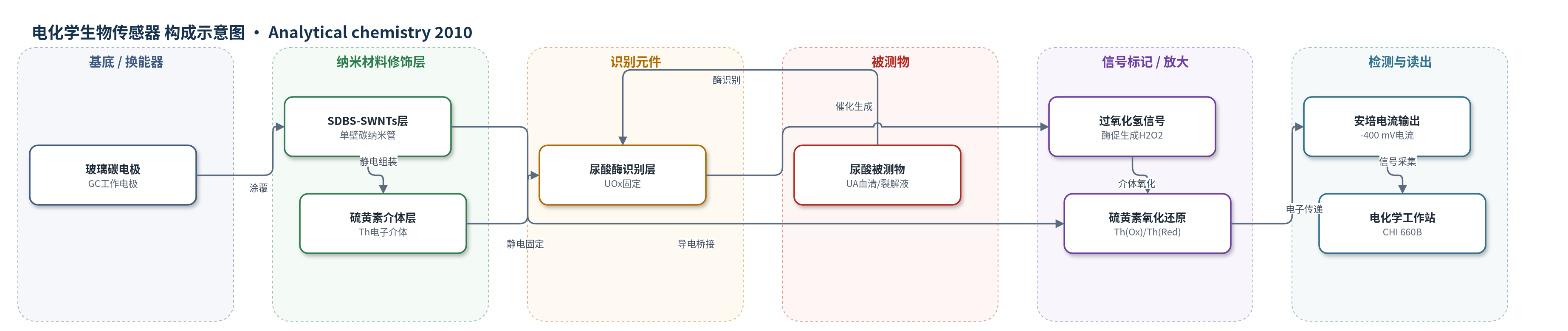
点击图片查看大图 · 依据论文自动绘制
传感器类型
电化学生物传感器
检测对象
尿酸(uric acid, UA);样品基质为血清(serum)和细胞裂解液(cell lysate,HEK 293A)
检测原理
尿酸酶(UOx)特异性识别并催化尿酸(UA)在氧气存在下氧化为别嘌呤(allantoin),同时生成过氧化氢(H2O2)和 CO2。生成的 H2O2 扩散至硫黄素(Th)-单壁碳纳米管(SWNTs)界面,被还原态硫黄素(Th(Red))化学还原为水,使硫黄素氧化为 Th(Ox);Th(Ox) 在电极表面接受电子再生为 Th(Red),形成可逆氧化还原循环。SWNTs 提供导电通道并加速电子转移,使 H2O2 还原在 -400 mV(vs SCE)低电位下发生,避免抗坏血酸、多巴胺等电活性干扰物氧化。UA 浓度越高,酶促生成 H2O2 越多,安培还原电流越大,从而实现定量检测。
检测灵敏度
LOD: 0.5 ± 0.05 µM;线性范围: 2 µM to 2 mM;slope: 6.5 ± 0.4 µA mM-1;sensitivity: ∼90 µA mM-1 cm-2;R^2 = 0.998
效应效果
该传感器在 -400 mV 下对 UA 选择性良好,葡萄糖(5 mM)、AA(0.1 mM)、DOPAC(0.1 mM)、DA(0.1 mM)、AP(0.1 mM)几乎无响应。响应时间约2 s。10个电极斜率 RSD 约2.4%,0.2 mM UA 10次测定 RSD 约2.8%。4℃储存1个月保留约90%;连续使用1周保留约85%,1个月约70%;-400 mV 极化2 h保留约92%。血清结果与医院参考值偏差 -4.6% 至 4.3%;HEK 293A 裂解液加标回收率 97.3%–104%,平均约100.9%、101.4%、101.1%,RSD 2.60%–3.43%。LOD 低于聚苯胺-UOx 的 10 µM,可用于生理和细胞内 UA 研究。
传感器的构成
- 基底/换能器电极:玻璃碳电极(GC, glassy carbon electrode)作为工作电极基底,提供电子转导界面。
- 纳米材料修饰层:单壁碳纳米管(SWNTs, single-walled carbon nanotubes)经十二烷基苯磺酸钠(SDBS)分散后涂覆于 GC 表面,形成导电网络并增大有效面积。
- 介体修饰层:硫黄素(Th, thionine)通过静电作用组装在 SDBS-SWNTs 表面,作为 H2O2 电催化还原介体并促进电子传递。
- 识别元件:尿酸酶(UOx, uricase)在 pH 7.4 下带负电,通过静电作用固定于带正电的 Th-SWNTs 表面,特异性催化 UA 氧化。
- 信号产物:过氧化氢(H2O2)由 UOx 催化 UA 氧化生成,在 -400 mV 下被 Th-SWNTs 电催化还原产生安培电流。
- 检测介质:0.1 M 磷酸盐缓冲液(PBS, pH 7.4)作为支持电解质;实际样品基质为血清和细胞裂解液。
- 电化学测量组件:饱和甘汞电极(SCE)作参比电极,铂丝作对电极,用于三电极安培/伏安测量。
中文摘要
本研究开发并验证了一种用于内源性(细胞裂解液)和生理性(血清)样品中尿酸(UA)测定的电化学方法。该方法基于尿酸酶-硫黄素-单壁碳纳米管/玻璃碳(UOx-Th-SWNTs/GC)生物传感器上对酶促生成过氧化氢(H2O2)的电催化还原,其中 Th-SWNTs 纳米结构同时作为电子介体和酶固定基质。通过将尿酸酶(UOx)固定于 Th-SWNTs 表面制备该生物传感器,其具有快速响应(约2 s)、低检出限(0.5 ± 0.05 µM)、宽线性范围(2 µM–2 mM)、高灵敏度(约90 µA mM−1 cm−2)以及良好的稳定性和重复性。由于采用低工作电位(-400 mV vs SCE),抗坏血酸、3,4-二羟基苯乙酸、4-乙酰氨基酚等常见干扰物不产生干扰。因此,本文展示了一个简单有效的传感平台,可在生理水平选择性检测 UA。该策略对于研究高血压、心血管疾病等状态下内源性和生理性 UA 的相对作用具有重要意义。
英文摘要
This work develops and validates an electrochemical approach for uric acid (UA) determinations in both endogenous (cell lysate) and physiological (serum) samples. This approach is based on the electrocatalytic reduction of enzymatically generated H(2)O(2) at the biosensor of uricase-thionine-single-walled carbon nanotube/glassy carbon (UOx-Th-SWNTs/GC) with the use of Th-SWNTs nanostructure as a mediator and an enzyme immobilization matrix. The biosensor, which was fabricated by immobilizing UOx on the surface of Th-SWNTs, exhibited a rapid response (ca. 2 s), a low detection limit (0.5 +/- 0.05 microM), a wide linear range (2 microM to 2 mM), high sensitivity (approximately 90 microA mM(-1) cm(-2)), as well as good stability and repeatability. In addition, the common interfering species, such as ascorbic acid, 3,4-dihydroxyphenylacetic acid, 4-acetamidophenol, etc., did not cause any interference due to the use of a low operating potential (-400 mV vs saturated calomel electrode). Therefore, this work has demonstrated a simple and effective sensing platform for selective detection of UA in the physiological levels. In particular, the developed approach could be very important and useful to determine the relative role of endogenous and physiological UA in various conditions such as hypertension and cardiovascular disease.