组成图示
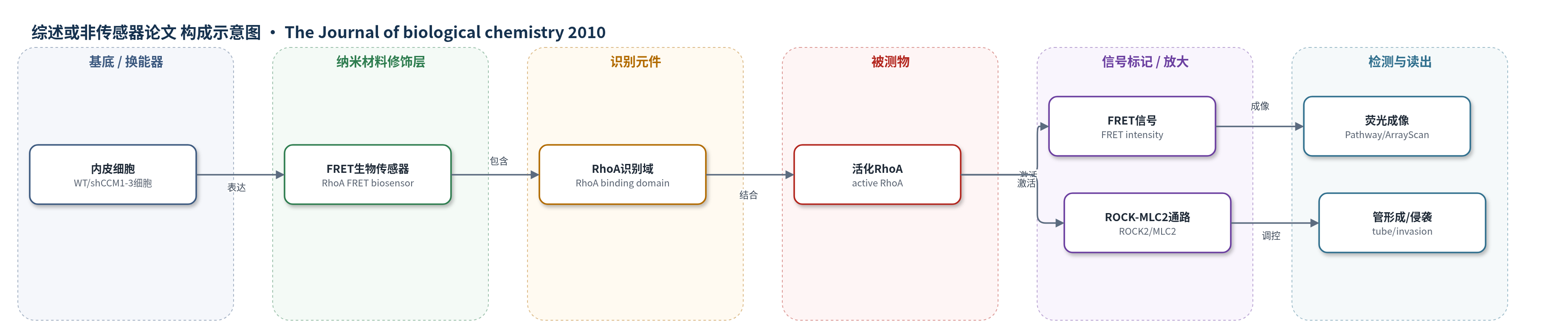
点击图片查看大图 · 依据论文自动绘制
传感器类型
综述或非传感器论文
检测对象
活化RhoA(active RhoA / RhoA-GTP);样品基质:活内皮细胞(WT 或 shCCM1/-2/-3 敲低细胞)
检测原理
该研究使用基于 FRET 的 RhoA 特异性生物传感器在活内皮细胞中检测 RhoA 活性。当 CCM1、CCM2 或 CCM3 缺失时,RhoA 蛋白表达升高并持续活化;活化 RhoA 与传感器识别域结合,引起传感器构象变化,使供体-受体间 FRET 效率升高。荧光成像系统采集 FRET 强度,细胞边缘、胞质和核内 FRET 信号增强反映 RhoA 活性升高。下游,活化 RhoA 激活 ROCK2,磷酸化 MLC2,改变肌动蛋白细胞骨架稳定性,抑制类血管管形成和细胞外基质侵袭;ROCK 抑制或 ROCK2 敲低可逆转该表型。
检测灵敏度
原文未报告LOD、线性范围、灵敏度斜率或R^2。
效应效果
该 FRET 生物传感器在活细胞中可定量 RhoA 活性空间分布。shCCM1、shCCM2 和 shCCM3 内皮细胞中 RhoA 表达分别升高约 23、37 和 28 倍;FRET 强度在胞质、核和细胞边缘分别较 WT 升高 1.62、2.33 和 1.48 倍(shCCM1),shCCM2/shCCM3 约升高 1.16–1.23 倍,差异具有统计学意义。ROCK2 敲低或 Y-27632 处理可挽救 CCM 敲低细胞的管形成和基质侵袭,其中 shROCK2 挽救效果优于 Y-27632,且 Y-27632 起效延迟。研究未报告实际样品回收率或与 ELISA/HPLC/qPCR 的对比,但将 ROCK 定位为 CCM 潜在治疗靶点。
传感器的构成
- 样品基质:内皮细胞(endothelial cells),包括 WT 和 shCCM1/-2/-3 敲低细胞,承载 RhoA 活性变化。
- 识别元件:RhoA 特异性 FRET 生物传感器(RhoA-specific FRET biosensor),用于响应活化 RhoA。
- 信号标记:FRET 荧光信号(FRET intensity,具体荧光蛋白未报道),RhoA 激活时 FRET 增强。
- 换能器/读出:荧光高内涵成像系统(Pathway、Cellomics ArrayScan),采集并伪彩色显示 FRET 强度。
- 对照/干预:shRNA 敲低 CCM1/-2/-3 或 ROCK2,以及 ROCK 抑制剂 Y-27632,用于验证 RhoA-ROCK 通路。
中文摘要
脑 cavernous malformation(CCM)是由 ccm1、ccm2 或 ccm3 失活突变引起的脑血管病变,可导致癫痫和卒中。本文报道,CCM1、CCM2 或 CCM3 蛋白表达缺失均使内皮细胞中 GTP 酶 RhoA 表达显著升高。利用 RhoA 特异性 FRET 生物传感器进行活细胞成像显示,CCM1、CCM2 或 CCM3 敲低后,RhoA 活性在细胞边缘、胞质和细胞核中均增加,其中 CCM1 缺失时胞质和核内 RhoA 激活尤为明显。增强的 RhoA 活性伴随 Rho 激酶(ROCK)依赖的肌球蛋白轻链 2(MLC2)磷酸化升高。功能上,CCM1、CCM2 或 CCM3 缺失抑制内皮细胞类血管管形成和细胞外基质侵袭,而 ROCK 化学抑制或 shRNA 敲低可挽救上述缺陷。研究首次界定 CCM1/2/3 在 CCM 病理中的 RhoA-ROCK 信号网络,并将 ROCK 确定为挽救内皮细胞功能的治疗靶点。
英文摘要
Cerebral cavernous malformations (CCM) are vascular lesions causing seizures and stroke. Mutations causing inactivation of one of three genes, ccm1, -2, or -3, are sufficient to induce vascular endothelial cell defects resulting in CCM. Herein, we show that loss of expression of the CCM1, -2, or -3 proteins causes a marked increase in expression of the GTPase RhoA. Live cell imaging with a RhoA-specific biosensor demonstrates increased RhoA activity with loss of CCM1, -2, or -3, with an especially pronounced RhoA activation in both the cytosol and the nucleus with loss of CCM1 expression. Increased RhoA activation was associated with Rho kinase-dependent phosphorylation of myosin light chain 2. Functionally, loss of CCM1, -2, or -3 inhibited endothelial cell vessel-like tube formation and extracellular matrix invasion, each of which is rescued by chemical inhibition or short hairpin RNA knockdown of Rho kinase. The findings, for the first time, define a signaling network for CCM1, -2, and -3 in CCM pathology, whereby loss of CCM1, -2, or -3 protein expression results in increased RhoA activity, with the activation of Rho kinase responsible for endothelial cell dysregulation. The results define Rho kinase as a therapeutic target to rescue endothelial cells from loss of CCM protein function.