组成图示
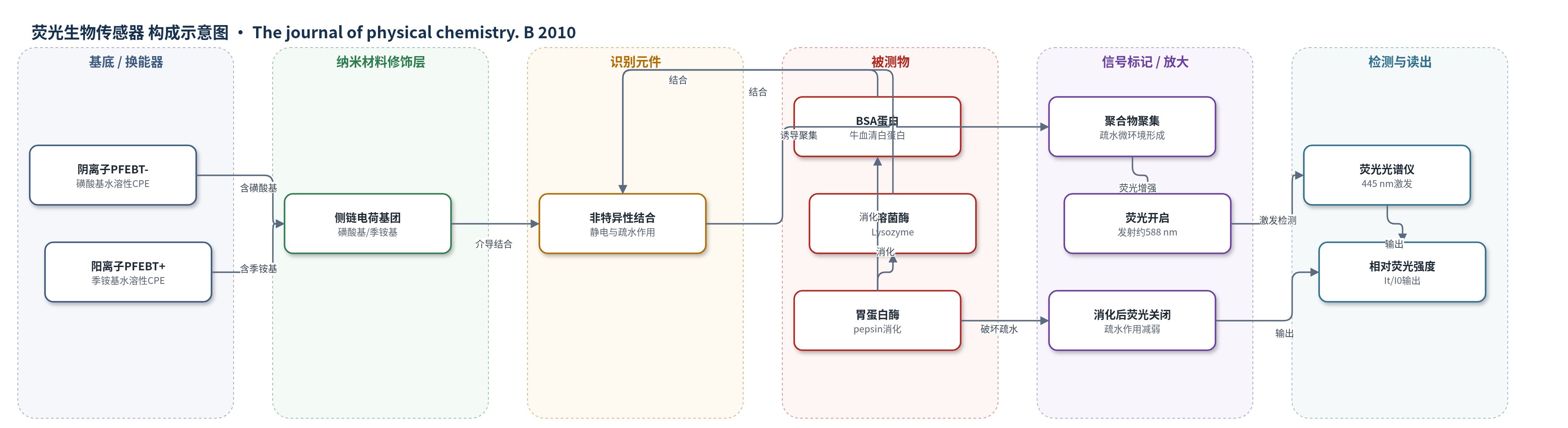
点击图片查看大图 · 依据论文自动绘制
传感器类型
荧光生物传感器
检测对象
牛血清白蛋白(BSA)、溶菌酶(Lysozyme);样品基质为15 mM PBS缓冲液(pH 7.4,pH效应实验用pH 2–14缓冲液)
检测原理
PFEBT的激发态具有电荷转移特征,在水溶液中非辐射衰减较强,荧光很弱。当BSA或溶菌酶加入后,蛋白与PFEBT通过静电吸引和/或疏水作用形成复合物,诱导聚合物链聚集并形成疏水微环境,减少水分子与π共轭主链接触,从而抑制电荷转移态的非辐射失活,使荧光强度增加且发射峰蓝移。随着蛋白浓度升高至0.8 µM,荧光增强趋于饱和。pH改变蛋白净电荷,可增强相反电荷间的静电吸引,使复合物更紧密,荧光进一步增加。胃蛋白酶消化破坏蛋白疏水结构,使疏水作用减弱,PFEBT/BSA复合物荧光下降,呈现light-off信号;而肽片段电荷密度与完整蛋白相近,静电作用保留,因此静电主导的复合物荧光下降较小。共轭聚合物聚集和激子迁移提供信号放大。
检测灵敏度
原文未报告LOD、线性范围、灵敏度斜率及相关系数。
效应效果
pH 7.4 PBS中,PFEBT+对BSA响应最强,0.8 µM时PL为466 au,增强17.3倍;PFEBT-对BSA为153 au,增强5.7倍。PFEBT-对溶菌酶增强约3.6倍,PFEBT+对溶菌酶无明显响应,体现电荷匹配选择性。pH调节可改变响应:PFEBT-/BSA在pH 2时强度由151 au升至273 au,λmax由588 nm蓝移至556 nm;PFEBT+/溶菌酶在pH 14时由28 au升至73 au,λmax移至565 nm。胃蛋白酶消化150 min后,PFEBT-/BSA荧光下降约70%,PFEBT-/溶菌酶约14%,PFEBT+/BSA约6%,说明疏水作用可被消化消除而静电作用保留。作者认为可用于无标记蛋白定量和蛋白酶活性监测。
传感器的构成
- 溶液相介质:15 mM PBS缓冲液(pH 7.4,pH效应实验用pH 2–14缓冲液),维持蛋白电荷状态并提供荧光测量环境
- 换能/信号材料:阴离子PFEBT-(poly[9,9-bis(4′-sulfonatobutyl)fluorenyleneethynylene-alt-4,7-(2,1,3-benzothiadiazole) disodium]),磺酸基赋予水溶性,与蛋白结合后聚集并荧光开启
- 换能/信号材料:阳离子PFEBT+(poly[9,9-bis(6′-(N,N,N-trimethylammonium)-hexyl)fluorenyleneethynylene-alt-4,7-(2,1,3-benzothiadiazole) dibromide]),季铵基赋予水溶性,与蛋白结合后聚集并荧光开启
- 修饰/识别基团:PFEBT侧链磺酸基(PFEBT-)与季铵基(PFEBT+),提供水溶性并介导静电相互作用
- 识别元件:PFEBT侧链电荷基团与蛋白表面电荷及疏水区域,通过静电吸引和疏水作用实现非特异性结合
- 信号标记物:PFEBT自身BT单元荧光(发射约588 nm),无外源荧光标记
- 消化调控元件:胃蛋白酶(pepsin),用于切割蛋白以削弱疏水作用并产生荧光关闭信号
- 信号读出:荧光光谱仪(Perkin-Elmer LS-55,445 nm激发,588 nm发射)
中文摘要
本文设计并合成了阳离子和阴离子聚芴乙烯基-苯并噻二唑共聚物(PFEBT),用于蛋白质的荧光开启检测。由于激发态具有电荷转移特征,PFEBT在水溶液中荧光很弱,但聚合物聚集可增强其黄色荧光。与蛋白结合时,PFEBT表现出荧光增强而非猝灭。静电作用和疏水作用均可促进PFEBT/蛋白复合物形成并提高荧光,增强程度取决于聚合物和蛋白性质。通过调节溶液pH改变蛋白净电荷,可有效调控静电相互作用及荧光增量。胃蛋白酶消化实验表明,静电作用诱导的荧光增强在蛋白被切割为肽片段后仍难以显著降低;而主要由蛋白疏水性决定的疏水作用可被消化削弱,使复合物呈现荧光关闭信号。该研究展示了利用非特异性相互作用实现无标记蛋白传感的可行性,并为生物传感器设计提供重要启示。
英文摘要
Cationic and anionic poly(fluorenyleneethynylene-alt-benzothiadiazole)s (PFEBTs) are designed and synthesized via Sonagashira coupling reaction to show light-up signatures toward proteins. Due to the charge transfer character of the excited states, the fluorescence of PFEBTs is very weak in aqueous solution, while their yellow fluorescence can be enhanced by polymer aggregation. PFEBTs show fluorescence turn-on rather than fluorescence quenching upon complexation with proteins. Both electrostatic and hydrophobic interactions between PFEBTs and proteins are found to improve the polymer fluorescence, the extent of which is dependent on the nature of the polymer and the protein. Changes in solution pH adjust the net charges of proteins, providing an effective way to manipulate electrostatic interactions and in turn the increment in the polymer fluorescence. In addition, the effect of protein digestion on the fluorescence of polymer/protein complexes is probed. The results indicate that electrostatic interaction induced polymer fluorescence increase cannot be substantially reduced through cleaving protein into peptide fragments. In contrast, hydrophobic interactions, mainly determined by the hydrophobicity of proteins, can be minimized by digestion, imparting a light-off signature for the polymer/protein complexes. This study thus not only highlights the opportunities of exerting nonspecific interactions for protein sensing but also reveals significant implications for biosensor design.