组成图示
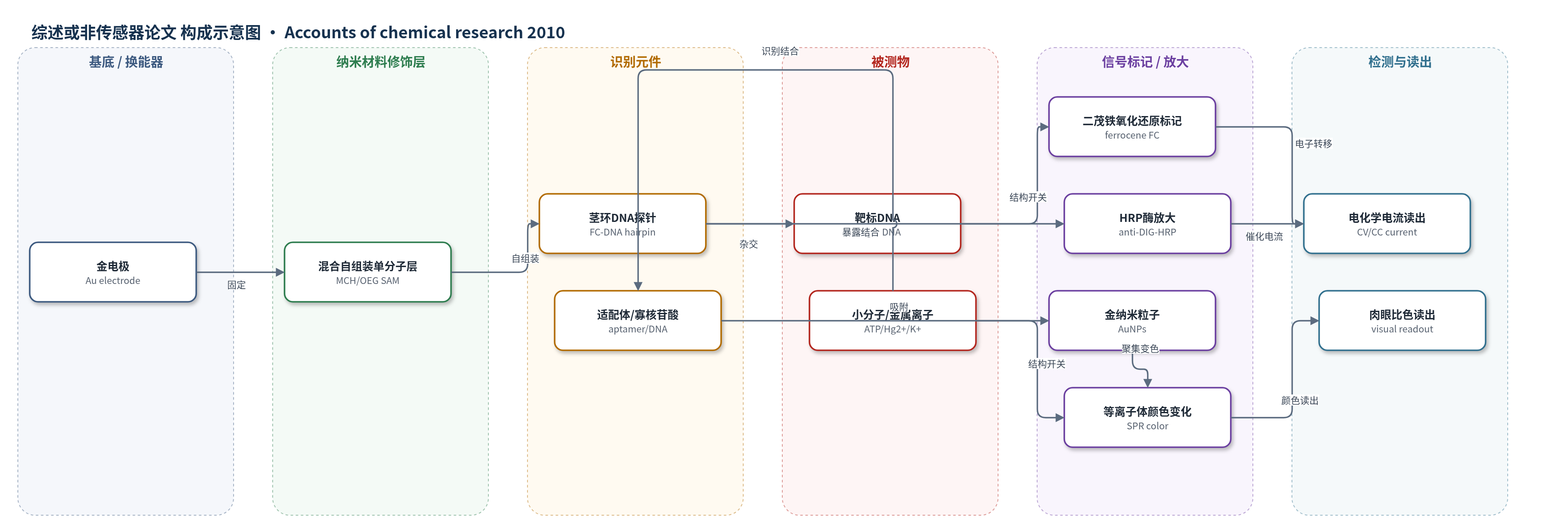
点击图片查看大图 · 依据论文自动绘制
传感器类型
综述或非传感器论文
检测对象
靶标DNA(target DNA,PCR产物/血清)、ATP(adenosine triphosphate,细胞裂解液)、可卡因(cocaine,掺假样品/生物流体)、汞离子(Hg2+,水样)、钾离子(K+,溶液)、pH(溶液)、肿瘤DNA标志物(P16/P21/P53,溶液)
检测原理
表面固定的茎环DNA或适配体探针在靶标结合后发生结构开关:靶标DNA杂交打开茎环,ATP或可卡因诱导适配体从双链/随机卷曲转为三级结构,富T DNA探针遇Hg2+形成刚性结构。这些变化改变二茂铁等氧化还原标记与金电极的距离,电子转移速率随距离指数变化,循环伏安或计时库仑电流随靶标浓度改变。酶型E-DNA中,杂交暴露DIG,抗DIG-HRP结合后HRP催化H2O2产生电化学电流,实现信号开启和放大。AuNP方案利用单链/双链或折叠/展开核酸对AuNP吸附、聚集的差异,靶标诱导AuNP聚集或解聚,SPR峰位移导致红-紫/蓝颜色变化;AuNP还可超猝灭荧光染料,靶标结合使染料远离AuNP而荧光开启。AuNP负载报告DNA或酶可进一步放大信号。
检测灵敏度
ATP传感器: LOD 10 nM;原始E-DNA: LOD ∼10 pM;酶型E-DNA: LOD 10 fM;AuNP计时库仑DNA传感器: LOD 10 fM
效应效果
E-DNA传感器可重复使用,适用于掺假样品和血清;ATP传感器不响应CTP、GTP、UTP,可直接检测细胞裂解液ATP,1 mM ATP产生最高10倍ON/OFF增益。酶型E-DNA可在百万倍非互补DNA存在下识别靶标并区分单碱基错配,还检测了大肠杆菌uidA基因PCR产物。OEG抗污表面抵抗蛋白吸附,在血清中保持性能。AuNP计时库仑传感器放大超过3个数量级,LOD 10 fM。AuNP比色法室温快速,工程化可卡因适配体数分钟内完成,较置换法约30 min更快;PDMS微流控芯片可无源肉眼检测Hg2+,适合现场即时检测。
传感器的构成
- 基底/换能器:金电极(Au electrode),用于电化学换能;PDMS微流控芯片,用于比色/肉眼读出
- 修饰层:混合自组装单分子层(SAM),含巯基DNA探针、巯基乙醇(MCH)或寡乙二醇(OEG)硫醇,用于固定探针、抑制非特异吸附并促进探针立起
- 识别元件:茎环DNA探针、互补DNA(cDNA)、抗ATP适配体、抗可卡因适配体、富T DNA探针、G-四联体探针、C-rich i-motif探针,用于识别靶标DNA、ATP、可卡因、Hg2+、K+等
- 信号标记物:二茂铁(ferrocene, FC)、地高辛(DIG)、生物素(biotin)、[Ru(NH3)6]3+、荧光染料FAM/Cy5/Rox,用于电化学或荧光信号
- 放大元件:金纳米粒子(AuNPs)、链霉亲和素(streptavidin)、抗DIG-HRP抗体、辣根过氧化物酶(HRP)和H2O2,用于信号放大
- 读出方式:循环伏安(CV)、计时库仑(CC)、电化学发光(ECL)、比色/SPR颜色、荧光或肉眼观察
中文摘要
本文综述了基于靶标响应结构开关的核酸生物传感器。作者指出,将配体诱导的DNA或适配体构象变化与电化学、光学换能器耦合,可构建无需标记、高选择性的传感平台。在电化学体系中,表面固定的茎环DNA或适配体探针带有二茂铁等氧化还原标记,靶标结合后改变标记物与金电极的距离,电子转移速率随之变化,从而通过循环伏安或计时库仑电流读出信号;引入混合自组装单分子层、OEG抗污表面、酶催化或金纳米粒子放大可进一步提高灵敏度。在光学体系中,金纳米粒子对单链/双链或折叠/展开核酸的吸附与聚集行为不同,靶标诱导的结构转换引起表面等离子体颜色变化或荧光猝灭/开启,可用于小分子和金属离子检测。作者还介绍了微流控芯片实现肉眼汞离子检测,并展望了拓展分析物谱和集成纳米材料的前景。
英文摘要
Interest in the development of sensitive, selective, rapid, and cost-effective biosensors for biomedical analysis, environmental monitoring, and the detection of bioterrorism agents is rapidly increasing. A classic biosensor directly transduces ligand-target binding events into a measurable physical readout. More recently, researchers have proposed novel biosensing strategies that couple ligand-induced structural switching of biomolecules with advanced optical and electronic transducers. This approach has proven to be a highly general platform for the development of new biosensors. In this Account, we describe a series of electrochemical and optical nucleic acid sensors that use target-responsive DNA structures. By employing surface-confined DNA structures with appropriate redox labels, we can monitor target-induced structural switching of DNA or aptamer-specific small molecule probes by measuring electrochemical currents that are directly associated with the distance between the redox label and the electrode surface. We have also demonstrated significant improvements in sensing performance through optimization of the DNA self-assembly process at electrode surfaces or the introduction of nanomaterial-based signal amplification. Alternatively, gold nanoparticles interact differently with folded and unfolded DNA structures, which provides a visual method for detecting target-induced structural switching based on the plasmonic change of gold nanoparticles. This novel method using gold nanoparticles has proven particularly suitable for the detection of a range of small-molecule targets (e.g., cocaine) and environmentally toxic metal ions (e.g., Hg(2+)). Rational sequence design of DNA aptamers improves the sensitivity and increases the reaction kinetics. Recently, we have also designed microfluidic devices that allow rapid and portable mercury detection with the naked eye. This Account focuses on the use of bulk and nanoscale gold and DNA/aptamer molecules. We expect that researchers will further expand the analyte spectrum and improve the sensitivity and selectivity of nucleic acid sensors using functional biomolecules, such as DNAzymes, peptide aptamers and engineered proteins, and nanomaterials of different sizes, dimensions and compositions, such as carbon nanotubes, graphene, silicon nanowires, and metal nanoparticles or nanorods.