组成图示
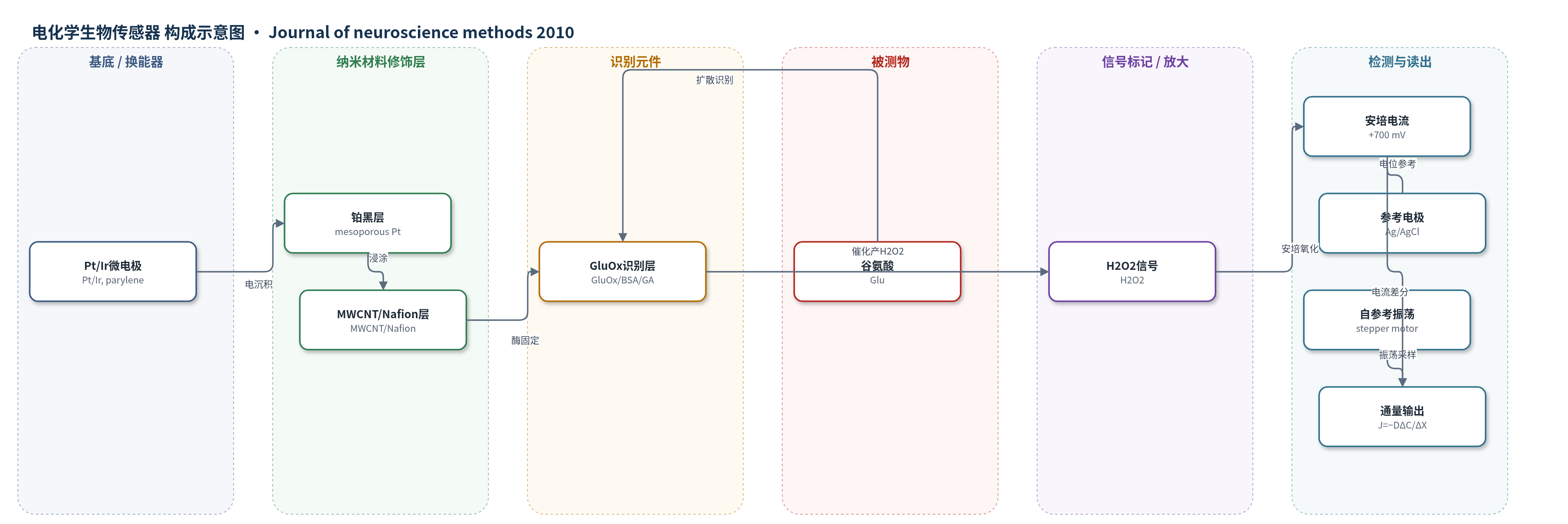
点击图片查看大图 · 依据论文自动绘制
传感器类型
电化学生物传感器
检测对象
谷氨酸(glutamate, Glu);样品基质:体外分化 P19 神经细胞培养体系(Locke’s buffer,细胞表面附近微环境)
检测原理
谷氨酸扩散至 GluOx 识别层后,被谷氨酸氧化酶催化氧化:glutamate + H2O → α-ketoglutarate + H2O2。生成的 H2O2 在 +700 mV 工作电位下于铂黑/MWCNT 修饰电极表面发生安培氧化,产生与 H2O2 浓度成正比的电流,从而间接反映局部谷氨酸浓度。铂黑与 MWCNT 增强电子转移,提高灵敏度。自参考模式下,微电极由步进电机在细胞表面附近两个固定位置之间振荡,连续记录两位置浓度差 ΔC 与固定位移 ΔX,依据 Fick 第一定律 J=−DΔC/ΔX 计算通量。相位敏感检测使同相漂移和噪声在差分中抵消,因此可非侵入地量化谷氨酸释放/摄取的方向、速率与大小,而不依赖复杂数学模型。
检测灵敏度
LOD: 0.9 ± 0.3 μM;线性范围: 0.1–100 μM(动态范围 0–130 μM);灵敏度: 0.5 nA μM−1(三电极)、46.6 pA μM−1(两电极自参考)、473 ± 57 μA μM−1 cm−2;R^2 = 0.97
效应效果
谷氨酸校准 R^2=0.97,自参考灵敏度 46.6 pA μM−1,t95=1.1±0.3 s。谷氨酸/抗坏血酸斜率比 275:1;稳定性≥14 天,灵敏度变化<5%。动态效率 ε=0.98(梯度)/0.94(细胞)。基础谷氨酸 5.8±0.6 μM,与微透析及 GluOx 微电极相近。钾刺激后流出 1.5±0.2 s 内出现,摄取持续 1.6±0.2 min;TBOA 抑制后峰值流出 370±86、峰值流入 54.1±1.3 fmol cm−2 s−1,低于未抑制 500±86、135±14 fmol cm−2 s−1,适用于动态神经递质转运研究。
传感器的构成
- 微电极基底:Pt/Ir 丝(2–5 μm 尖端,聚对二甲苯绝缘)作为工作电极骨架
- 铂黑修饰层:mesoporous Pt 电沉积层,提高导电性与电催化活性
- 纳米修饰层:MWCNT/Nafion 浸涂层,增强电子转移并辅助固定酶
- 识别元件:GluOx(谷氨酸氧化酶)通过 1% BSA、0.125% glutaraldehyde 与 1% GluOx 混合液浸涂固定,特异性氧化谷氨酸
- 固定/封闭层:BSA 与 glutaraldehyde 形成交联固定基质,稳定 GluOx
- 信号产物:H2O2(GluOx 催化谷氨酸氧化产生),在 +700 mV 下被安培检测
中文摘要
神经递质转运动力学的定量研究受限于缺乏能在生理条件下直接监测生物活性通量的工具。传统神经递质释放/摄取研究依赖非选择性电记录推断,具有侵入性/破坏性,且时间分辨率不足。电化学生物传感器的发展提高了生理与病理条件下神经递质浓度的检测能力,酶传感器结合碳纳米管等性能增强材料是重要方向。然而,由于灵敏度较低、漂移与噪声较大、信噪比不足,以及难以量化突触传递中的快速神经化学动力学,这些技术尚未成为主流神经科学研究工具。自参考技术可克服上述问题,实现生物物理转运的非侵入定量。本文报道一种自参考碳纳米管修饰谷氨酸氧化酶生物传感器,用于监测神经/神经元细胞附近谷氨酸通量。基础谷氨酸浓度与其他体内、体外测量结果相近。该传感器以自参考(振荡)模式测量电刺激期间神经细胞附近的净谷氨酸通量:刺激前平均流入为33.9±6.4 fmol cm−2 s−1;刺激后立即出现谷氨酸流出,随后出现50–150 fmol cm−2 s−1范围的摄取。使用threo-β-benzyloxyaspartate抑制摄取后,重复细胞平均表面通量(1.1±7.4 fmol cm−2 s−1)显著低于未抑制细胞。该技术对研究动态生理条件下与神经传递相关的神经病理状态具有重要价值。
英文摘要
Quantification of neurotransmitter transport dynamics is hindered by a lack of sufficient tools to directly monitor bioactive flux under physiological conditions. Traditional techniques for studying neurotransmitter release/uptake require inferences from non-selective electrical recordings, are invasive/destructive, and/or suffer from poor temporal resolution. Recent advances in electrochemical biosensors have enhanced in vitro and in vivo detection of neurotransmitter concentration under physiological/pathophysiological conditions. The use of enzymatic biosensors with performance enhancing materials (e.g., carbon nanotubes) has been a major focus for many of these advances. However, these techniques are not used as mainstream neuroscience research tools, due to relatively low sensitivity, excessive drift/noise, low signal-to-noise ratio, and inability to quantify rapid neurochemical kinetics during synaptic transmission. A sensing technique known as self-referencing overcomes many of these problems, and allows non-invasive quantification of biophysical transport. This work presents a self-referencing CNT modified glutamate oxidase biosensor for monitoring glutamate flux near neural/neuronal cells. Concentration of basal glutamate was similar to other in vivo and in vitro measurements. The biosensor was used in self-referencing (oscillating) mode to measure net glutamate flux near neural cells during electrical stimulation. Prior to stimulation, the average influx was 33.9+/-6.4 fmol cm(-2)s(-1)). Glutamate efflux took place immediately following stimulation, and was always followed by uptake in the 50-150 fmol cm(-2)s(-1) range. Uptake was inhibited using threo-beta-benzyloxyaspartate, and average surface flux in replicate cells (1.1+/-7.4 fmol cm(-2)s(-1)) was significantly lower than uninhibited cells. The technique is extremely valuable for studying neuropathological conditions related to neurotransmission under dynamic physiological conditions.