组成图示
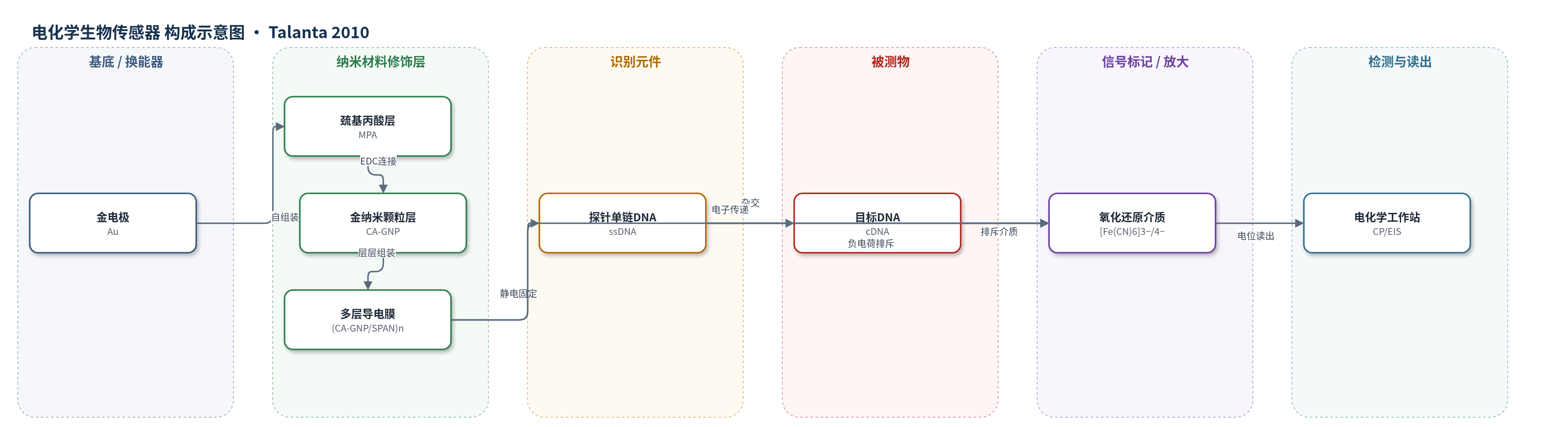
点击图片查看大图 · 依据论文自动绘制
传感器类型
电化学生物传感器
检测对象
目标DNA(target DNA/cDNA,PAT基因20碱基片段);样品基质:2× SSC杂交缓冲液(pH 7.0)
检测原理
该传感器采用无标记电位法检测DNA杂交,未使用酶催化或核酸扩增。带负电的探针ssDNA通过静电作用固定在(CA-GNP/SPAN)n多层膜外层CA-GNP上;目标DNA加入后与探针杂交形成双链DNA,使电极界面负电荷密度增加。界面负电荷排斥溶液中的[Fe(CN)6]3−/4−氧化还原介质接近导电膜,导致电子转移阻力增大。SPAN纳米纤维提供自掺杂导电通路,CA-GNP促进电荷跨膜转移,使介质电子转移变化可被电极感知。恒电流计时电位法施加恒定电流,过电位ΔE=IR′随总电极电阻R′增大而升高;电化学阻抗谱中半圆直径对应电子转移电阻Ret,也随杂交程度增大。ΔR′或ΔRet与目标DNA浓度对数呈线性关系,从而实现定量检测。
检测灵敏度
LOD: 2.13 × 10⁻¹³ mol/L(CP,3σ);LOD: 2.34 × 10⁻¹³ mol/L(EIS,3σ);线性范围: 1.0 × 10⁻¹² mol/L–1.0 × 10−7 mol/L;R = 0.9899(CP);R = 0.9931(EIS);斜率: 177.14 Ω/decade(CP);斜率: 204.91 Ω/decade(EIS)。
效应效果
该传感器对完全互补目标DNA响应明显,非互补DNA响应可忽略,单碱基和双碱基错配DNA响应较小,可区分错配序列。探针电极在0.1 mol/L NaOH、HCl、Tris–HCl(pH 7.0)和2× SSC(pH 7.0)中浸泡60 min后过电位无下降;(CA-GNP/SPAN)3膜在B–R缓冲液(pH 7.0)放置40 h后峰电流下降约10%,随后至少10 d无进一步下降。检测1.0×10⁻¹⁰ mol/L目标DNA时,5个平行电极ΔE为238、234、232、228、226 mV,RSD为2.82%(n=5)。沸水10 min变性后可再生并重复5次不损失灵敏度。CP测量30 s内完成,快于EIS,可用于PAT基因快速定性检测。
传感器的构成
- 基底/换能器电极:金电极(Au electrode),经清洗、抛光和电化学活化,提供导电基底与电子转移动力。
- 自组装前驱层:3-巯基丙酸(MPA)自组装膜,在金表面形成稳定界面,其羧基经EDC活化后用于连接CA-GNP。
- 共价稳定层:半胱胺包覆金纳米颗粒(CA-GNP),通过EDC与MPA共价沉积,形成稳定导电界面并提供氨基/正电荷位点。
- 多层导电膜:磺化聚苯胺纳米纤维(SPAN)与CA-GNP交替层层组装为(CA-GNP/SPAN)n,SPAN提供自掺杂导电性,CA-GNP稳定SPAN并促进电荷转移。
- 识别元件:探针单链DNA(ssDNA),带负电磷酸骨架通过静电亲和固定在外层CA-GNP上,用于特异性识别目标DNA。
- 信号介质:[Fe(CN)6]3−/4−氧化还原电对,作为无标记检测的氧化还原指示剂,其接近电极的电子转移受DNA负电荷影响。
- 读出装置:电化学工作站(CHI 660C),通过恒电流计时电位法(CP)和电化学阻抗谱(EIS)记录过电位、总电阻或电子转移电阻。
中文摘要
本文报道了一种基于磺化聚苯胺(SPAN)纳米纤维与半胱胺包覆金纳米颗粒(CA-GNP)层层组装膜的电化学DNA生物传感器,用于核酸的快速、特异性无标记检测。首先在金电极表面自组装3-巯基丙酸(MPA)前驱膜,并通过EDC活化将CA-GNP共价沉积于Au/MPA电极,形成稳定导电基底;随后利用静电作用将SPAN纳米纤维与CA-GNP交替层层组装为(CA-GNP/SPAN)n膜。循环伏安法以[Fe(CN)6]3−/4−为氧化还原指示剂监测多层膜生长,该膜在中性介质中电子转移良好。带负电的探针单链DNA(ssDNA)通过静电亲和固定在外层CA-GNP上。恒电流计时电位法(CP)和电化学阻抗谱(EIS)以溶液中的[Fe(CN)6]3−/4−为介质,用于探针固定与杂交的电化学读出;EIS表征电子转移电阻,CP提供界面总电阻,二者与导电支撑上的电子转移电阻具有良好相关性。CP可在数秒内完成,适合作为快速换能手段。基于(CA-GNP/SPAN)n膜,20碱基目标DNA的检测限为2.13×10−13 mol/L,并展示了检测错配DNA的可行性。
英文摘要
This paper described a novel electrochemical DNA biosensor for rapid specific detection of nucleic acids based on the sulfonated polyaniline (SPAN) nanofibre and cysteamine-capped gold nanoparticle (CA-G(NP)) layer-by-layer films. A precursor film of 3-mercaptopropionic acid (MPA) was firstly self-assembled on the Au electrode surface. CA-G(NP) was covalently deposited on the Au/MPA electrode to obtain a stable substrate. SPAN nanofibre and CA-G(NP) were alternately layer-by-layer assembled on the stable substrate by electrostatic force. Cyclic voltammetry was used to monitor the consecutive growth of the multilayer films by utilizing [Fe(CN)(6)](3-/4-) as the redox indicator. The (CA-G(NP)/SPAN)(n) films showed satisfactory ability of electron transfer and excellent redox activity in neutral media. Negatively charged probe ssDNA was immobilized on the outer layer of the multilayer film (CA-G(NP)) through electrostatic affinity. Chronopotentiometry and electrochemical impedance spectroscopy were employed to obtain the direct electrochemical readout for probe ssDNA immobilization and hybridization using [Fe(CN)(6)](3-/4-) in solution as the mediator. While electrochemical impedance spectroscopy led to the characterization of the electron-transfer resistance at the electrode, chronopotentiometry provided the total resistance at the interfaces of the modified electrodes. A good correlation between the total electrode resistances and the electron-transfer resistances at the conducting supports was found. Chronopotentiometry was suggested as a rapid transduction means (a few seconds). Based on the (CA-G(NP)/SPAN)(n) films, the target DNA with 20-base could be detected up to 2.13x10(-13)mol/L, and the feasibility for the detection of base-mismatched DNA was also demonstrated.