组成图示
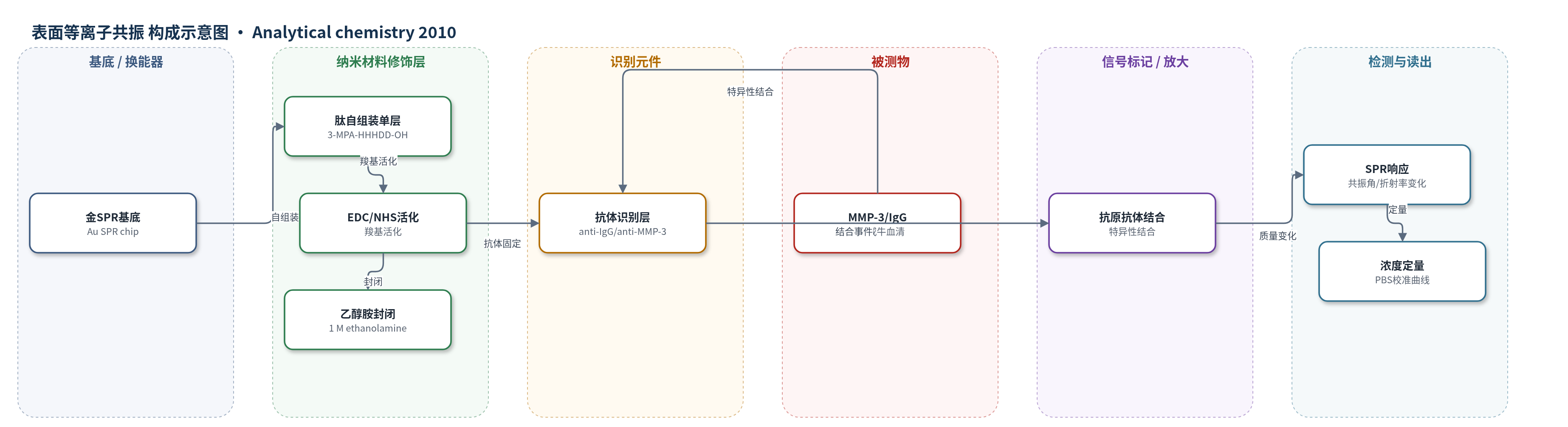
点击图片查看大图 · 依据论文自动绘制
传感器类型
表面等离子共振(SPR)生物传感器
检测对象
免疫球蛋白G(IgG)、基质金属蛋白酶-3(MMP-3);样品基质:PBS、未稀释牛血清(bovine serum)
检测原理
传感器以金SPR芯片为基底,3-MPA-HHHDD-OH肽通过N端巯基在金表面形成自组装单层,C端Asp羧基暴露。EDC/NHS将羧基活化为NHS酯,共价固定抗IgG或抗MMP-3抗体,乙醇胺封闭剩余位点。当PBS或牛血清中的IgG/MMP-3流过时,目标蛋白与抗体特异性结合,使SPR界面质量与局部折射率增加,引起共振角或波长偏移。响应大小随结合量增加而增大,在低浓度区近似线性,高浓度区受Langmuir等温线饱和影响。该设计无信号放大,依靠低污损肽单层抑制血清蛋白非特异吸附,使PBS校准可直接用于未稀释血清定量。
检测灵敏度
LOD: 1-10 pM(IgG,PBS);LOD: 3 pM(IgG低灵敏度区,图5);LOD: 0.11 nM(IgG高灵敏度区);LOD: 0.14 nM(MMP-3,PBS);线性范围: 0.5-50 nM(MMP-3);R^2 = 0.96(MMP-3);3σ = 0.03 nm(IgG LOD计算)
效应效果
3-MPA-HHHDD-OH单层对牛血清非特异吸附为32±5 ng/cm2,低于PEG约100 ng/cm2和CM-Dextran 829±46 ng/cm2;空白血清响应可忽略,表明抗干扰和稳定性良好。IgG传感器PBS检测限1-10 pM,优于或接近SERS、SPR、化学发光、ASV和LSPR等多步方法。MMP-3传感器PBS中LOD 0.14 nM,线性至50 nM;25 nM加标血清预测浓度32±2 nM,PBS预测30±2 nM,12.5 nM血清预测17±1 nM,1:1 PBS/血清预测12±4 nM,说明血清基质影响小。方法无需样品前处理、二抗或信号放大,适合复杂生物基质直接检测。
传感器的构成
- 基底/换能器:金表面SPR芯片(Au SPR chip),提供表面等离子共振换能界面
- 抗污修饰层:3-MPA-HHHDD-OH二元图案化肽自组装单层(SAM),N端3-MPA结合金,C端Asp羧基暴露,降低非特异吸附
- 活化层:EDC/NHS,活化肽C端羧基形成NHS酯,用于共价固定抗体
- 识别元件:抗人IgG或抗人MMP-3抗体(anti-human IgG / anti-human MMP-3),特异性结合目标蛋白
- 封闭剂:1 M乙醇胺(ethanolamine,pH 8.5),封闭未反应位点
- 样品基质:PBS或未稀释牛血清(bovine serum),提供IgG或MMP-3
- 信号读出:SPR仪器(Kretchmann/Dove prism setup),记录结合引起的共振响应变化
中文摘要
本文报道了一种基于二元图案化肽自组装单层(SAM)的表面等离子共振(SPR)生物传感器,用于在未经处理的粗牛血清中直接检测基质金属蛋白酶-3(MMP-3)。作者筛选了多种3-MPA-Ax-By-OH肽,发现3-MPA-HHHDD-OH在抗非特异性吸附方面优于其他肽及聚乙二醇(PEG)。该肽N端以3-巯基丙酸(3-MPA)结合金表面,C端保留羧基,可经EDC/NHS化学共价固定抗体。基于该单层的IgG传感器在PBS中检测限为1-10 pM,与常见光学或电化学方法相当。进一步以抗MMP-3抗体构建传感器,PBS中MMP-3检出限为0.14 nM,线性范围可达50 nM;使用PBS校准曲线即可在未稀释牛血清中低纳摩尔定量MMP-3,且血清响应与PBS统计一致,空白血清非特异吸附可忽略。结果表明,二元图案化肽SAM适用于复杂生物基质中的直接生物传感。
英文摘要
Near-zero fouling monolayers based on binary patterned peptides allow low nanomolar detection of the matrix metalloproteinase-3 (MMP-3) directly in crude bovine serum, without sample pretreatment, secondary antibody detection or signal amplification. The peptide 3-MPA-HHHDD-OH (3-MPA, 3-mercaptopropionic acid) was found optimal compared to other binary patterned peptides based on 3-MPA-A(x)-B(y)-OH, where 0 <or= x, y <or= 5, and x + y = 5, and compared to PEG. In this study, amino acid A was His, Asp, Ser, or Leu, and amino acid B was His, Asp, or Ser. Zwitterionic peptides and other peptides exhibited excellent resistance to nonspecific adsorption. Binary patterned peptides were capped with 3-MPA on the N-terminus providing a monolayer with the C-terminus carboxylic acid available to subsequently immobilize antibodies. Thereby, an IgG biosensor demonstrated the efficiency of binary patterned peptides in SPR biosensing with a detection limit of 1-10 pM in PBS, similar to other optical or electrochemical techniques. This protocol was applied to establish a calibration curve for MMP-3, an analyte of clinical interest for many pathologies and a potential indicator of cancer. The LOD for MMP-3 was 0.14 nM in PBS, with a linearity of up to 50 nM. With the use of PBS calibration, MMP-3 was quantified at low nanomolar in undiluted bovine serum. The SPR response in serum was statistically the same as in PBS. A sensor exposed to blank serum exhibited negligible nonspecific adsorption. Hence, binary patterned peptides are suitable for biosensing directly in complex biological matrixes.