组成图示
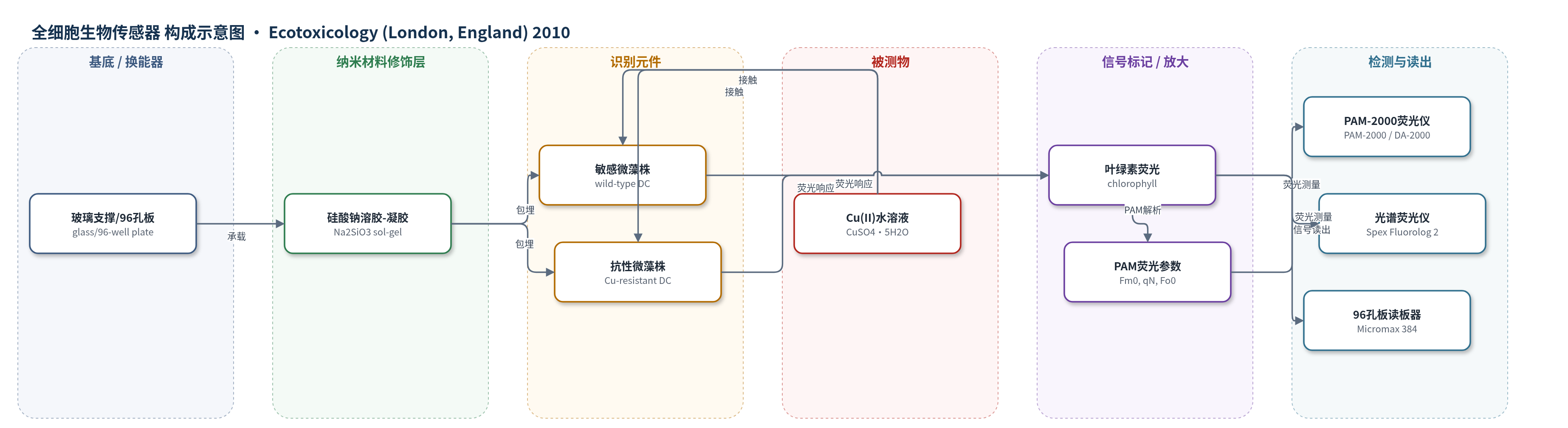
点击图片查看大图 · 依据论文自动绘制
传感器类型
全细胞生物传感器
检测对象
二价铜离子(Cu(II), copper(II)),样品基质为水样(水库水、饮用水/供水,实验用 BG-11 培养基)
检测原理
固定化微藻作为全细胞识别元件,Cu(II) 作用于藻细胞后干扰光系统II(PSII)光合电子传递,主要影响氧化侧及反应中心,使激发能非光化学耗散增加。PAM 以弱测量光脉冲和强饱和脉冲交替照射藻膜,分别获得开放/关闭 PSII 反应中心时的最小与最大叶绿素荧光(Fo0、Fm0),并计算光化学淬灭系数 qP、非光化学淬灭系数 qN 和有效量子产额 Y。Cu(II) 浓度升高时,Fm0 下降、qN 上升,qN 对低浓度铜最敏感,经四参数 logistic 曲线拟合实现定量。敏感野生型与 Cu 抗性突变株响应差异用于增强选择性,无需外源标记或酶放大。
检测灵敏度
LOD: 0.6 mg l-1;LOD: 0.65 ppm;动态范围: 1.1–8.4 mg l-1;EC50: 3.1 mg l-1;r > 0.995
效应效果
该方法以 PAM 饱和脉冲法优于普通光谱荧光法:流动池荧光信号随铜浓度先升后降,不能定量;96孔板法需 48 h 才出现明显信号,灵敏度不足。PAM 法中 qN 和 Fm0 最敏感,75 min 接触即可筛查。野生型 DC 的 qN 对 Cu(II) 的 LOD 为 0.6 mg/L(结果中为 0.65 ppm),动态范围 1.1–8.4 mg/L,EC50 为 3.1 mg/L,低于饮用水 2 mg/L 限值。加入法规限值水平的 Cr(50 μg/L)、Hg(1.0 μg/L)和 simazin(0.1 μg/L)后,qN 变化与单独铜相近,显示抗干扰能力。敏感株与耐受 1.9 mg/L Cu(II) 的抗性株响应不同,可增强选择性;作者认为该双株 PAM 生物传感器可用于水样铜超标现场筛查。
传感器的构成
- 基底/支撑层:玻璃支撑(glass supports)或96孔玻璃底板(96-well glass bottom plates),承载溶胶-凝胶膜并适配光纤或读板器。
- 固定化基质层:硅酸钠(Na2SiO3)溶胶-凝胶(sol-gel),以 HCl 酸化交联,甘油(glycerol)改善生物相容性并减少干燥收缩。
- 识别/响应元件:Dictyosphaerium chlorelloides(DC)微藻全细胞,野生型敏感株与 Cu 抗性突变株,Cu(II) 抑制 PSII 引起荧光参数变化。
- 被测物接触层:Cu(II) 水溶液(CuSO4·5H2O 配制),通过流动池或96孔板与藻膜接触。
- 信号产生层:藻体内叶绿素(chlorophyll)荧光,PAM 脉冲饱和法读取 Fo0、Fm0、qN 等,无需外源标记物。
- 读出装置:PAM-2000 便携式叶绿素荧光仪(Heinz Walz)或 Spex Fluorolog 2 光谱荧光仪(Jovin-Ivon)配光纤/96孔板读板器。
中文摘要
为监测水库和饮用水中铜浓度,将绿藻 Dictyosphaerium chlorelloides 细胞包埋于硅酸盐溶胶-凝胶基质中,比较光谱荧光法与脉冲幅度调制(PAM)荧光法对 Cu(II) 毒性响应的检测性能。光谱荧光法采用光纤耦合流动池或96孔板读板器测量叶绿素荧光;PAM法采用便携式叶绿素荧光仪和脉冲饱和法测定 Fo0、Fm0、Y、qP、qN 等参数。结果表明,Fm0 和 qN 对铜浓度变化最敏感,其中 qN 在75 min接触后提供适合水样筛查的信号。D. chlorelloides PAM 生物传感器对 Cu(II) 的检出限为 0.6 mg/L,低于饮用水法规限值,可用于判断铜是否超标。作者还通过逐级诱导获得耐受 1.9 mg/L (30 μM) Cu(II) 的抗性藻株,以增强铜测定的选择性,为构建敏感/抗性双株铜生物传感器提供基础。
英文摘要
Test methods are needed to monitor Cu concentrations in reservoirs and water supplies. Dictyosphaerium chlorelloides (Chlorophyta) cells were immobilized in a silicate sol-gel and the toxic effects of Cu(II) were examined using different techniques: fluorescence measurements (using a spectrofluorometer with an optic fiber coupled to a flow cell or a 96-well-plate reader) or by Pulse Amplitude Modulation (PAM) parameters using a portable instrument and the pulse saturation method. Fm' and qN were the most sensitive indicator parameters when performing Cu analysis in water. D. chlorelloides PAM biosensor presented a detection limit of 0.6 mg l(-1) for Cu(II), within the limits to establish if Cu concentrations exceeded regulatory levels. Moreover, a 1.9 mg Cu l(-1) (30 microM) resistant strain of the D. chlorelloides microalgae was produced in order to obtain more selectivity on the metal determination.