组成图示
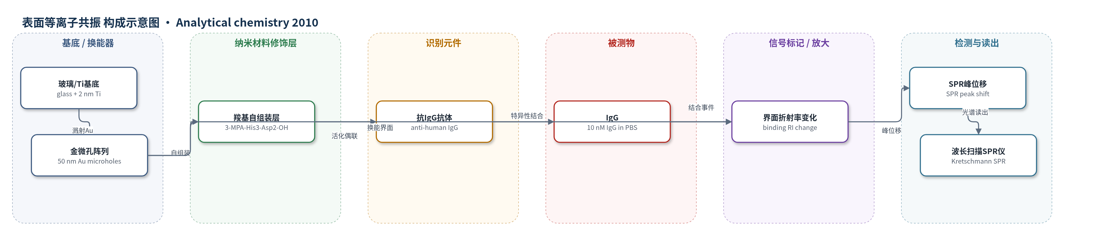
点击图片查看大图 · 依据论文自动绘制
传感器类型
表面等离子共振(SPR)生物传感器
检测对象
免疫球蛋白G(IgG),样品基质为 PBS(磷酸盐缓冲液)
检测原理
传感器以金微孔阵列作为 SPR 换能层,表面先自组装 3-MPA-(histidine)3-(aspartic acid)2-OH 单分子层,再用 EDC/NHS 活化羧基并共价偶联抗人 IgG 抗体。当 PBS 中的 IgG 与表面抗体特异性结合时,金/溶液界面局部质量与折射率增加,改变传播表面等离子体的共振条件。在 Kretschmann 棱镜激发下,P 偏振反射光谱中的 SPR 吸收峰发生波长位移,结合量越大,红移越明显。微孔阵列相比连续金膜具有更短的电磁场穿透深度,因此对近表面结合事件产生更强响应,实现无标记、实时检测。
检测灵敏度
RI 灵敏度: 2742 nm/RIU(连续膜);4200–2800 nm/RIU(微孔阵列);RI 分辨率: 1.5 × 10^-6 RIU
效应效果
作者以抗人 IgG 功能化表面检测 10 nM IgG:三角阵列响应 2.1±0.6 nm,微孔阵列 0.91±0.18 nm,连续金膜 0.69±0.11 nm。微孔阵列对 16-MHA 单分子层形成的响应约为连续膜 2 倍,折射率分辨率 1.5×10^-6 RIU,光谱噪声 0.0055 nm,与连续膜 0.0027 nm 相当,明显优于三角阵列 0.096 nm。微孔阵列兼具较高折射率灵敏度、较短穿透深度和与现有 SPR 仪器兼容的优点,作者认为其可替代连续金膜用于 SPR 生物传感。
传感器的构成
- 基底:玻璃盖玻片(glass coverslip),承载金膜并配合棱镜激发 SPR
- 粘附层:2 nm Ti,增强 Au 与玻璃结合
- 换能层:50 nm Au 微孔阵列/三角阵列(Au microhole arrays/triangles),Kretschmann 构型下激发传播 SPR
- 识别功能层:3-MPA-(histidine)3-(aspartic acid)2-OH 自组装单分子层,提供羧基用于 EDC/NHS 活化
- 识别元件:抗人 IgG 抗体(anti-human IgG),经 EDC/NHS 共价偶联,特异性结合 IgG
- 封闭剂:1 M 乙醇胺(ethanolamine, pH 8.5),封闭未反应 NHS 位点
- 读出系统:波长扫描 Kretschmann SPR 光谱仪,监测 P/S 偏振反射光谱中 SPR 峰位移
中文摘要
本文报道了利用改进纳米球光刻法在玻璃基底上制备微米级金三角阵列和微孔阵列,并在 Kretschmann 构型下研究其表面等离子共振(SPR)传感性能。采用直径 0.65、0.82、1.0、1.5 和 3.2 μm 的乳胶球作为掩模,获得边长 275–2000 nm 的金三角阵列以及不同周期、孔径和深度的微孔阵列。通过蔗糖溶液折射率标定、单分子层形成和动力学实验,系统考察了结构参数对折射率灵敏度、工作折射率范围、光谱峰宽、光谱噪声和折射率分辨率的影响。结果表明,孔径约为周期一半的微孔阵列兼具纳米颗粒局域表面等离子共振和薄膜 SPR 的优点,折射率灵敏度超过 3000 nm/RIU,对单分子层形成的响应较连续金膜提高约 2 倍,折射率分辨率达 10^-6 RIU。进一步以抗人 IgG 抗体功能化表面检测 10 nM IgG,微等离子体材料较连续金膜给出更强响应,显示其作为现有 SPR 仪器兼容的新型传感平台的潜力。
英文摘要
Metallic thin films patterned with micrometer size triangle or hole arrays present plasmonic properties when excited in the Kretschmann configuration, that are improved in comparison to conventional thin film surface plasmon resonance (SPR). These optical properties can be tuned by varying the physical aspects of the microplasmonic structures. Triangles and microhole arrays were prepared with modified nanosphere lithography (NSL) using latex spheres of 0.65, 0.82, 1.0, 1.5, or 3.2 microm in diameter. This allowed the preparation of triangles with edge lengths between 275 to 2000 nm and microhole arrays of various periodicities, diameters, and hole depths. These microstructures were studied to understand the relationship between the physical aspects and the optical properties, such as the sensitivity, working refractive index range, spectral width of the plasmonic peaks, spectral noise, and refractive index resolution. Microhole arrays with a hole diameter equal to half the periodicity were found to combine the advantages of both localized surface plasmon resonance (LSPR) on nanoparticles and SPR on a thin film. These microhole arrays exhibited high sensitivity to refractive index (>3000 nm/RIU), sensitivity to monolayer formation (2-fold improvement compared to thin films), and excellent refractive index resolution (10(-6) RIU). Finally, a biosensor for the detection of 10 nM of immunoglobulin G (IgG) exhibited a greater response with microplasmonic materials compared to conventional thin Au films. Hence, these novel plasmonic materials exhibit a strong potential as an SPR sensing platform. They can be implemented on existing instrumentation and use detection protocols developed for current SPR sensors.