组成图示
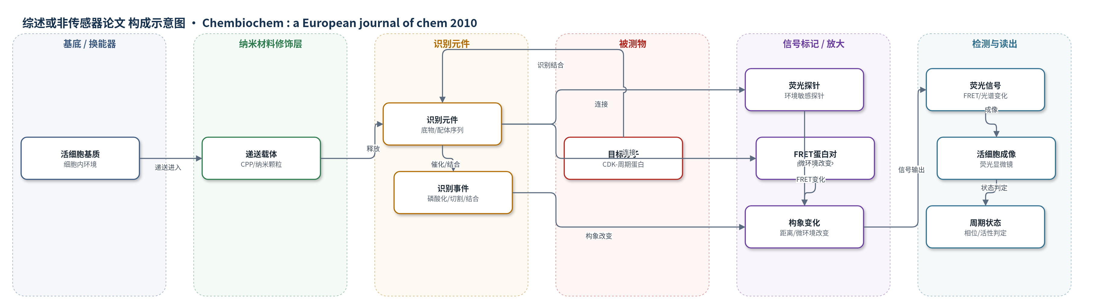
点击图片查看大图 · 依据论文自动绘制
传感器类型
综述或非传感器论文
检测对象
细胞周期调节因子:周期蛋白依赖性激酶(CDKs)、周期蛋白(cyclins)、Cdc25磷酸酶(Cdc25 phosphatases)、Plk1激酶(Plk1 kinase)、ATM激酶(ATM)、Aurora B激酶(Aurora B)、半胱天冬酶(caspases)、GTP酶(GTPases)、钙离子(Ca2+);样品基质:活细胞、肿瘤细胞/组织、细胞裂解液。
检测原理
综述所述荧光生物传感器通常由识别元件、报告序列和荧光标记组成。目标酶或配体与识别元件发生磷酸化、切割、结合或诱导构象变化后,识别域对修饰后的报告序列产生结合,或使环境敏感荧光探针的微环境改变;在FRET型传感器中,该变化使供体与受体自荧光蛋白(如EGFP/mRFP或CFP/YFP)距离或取向改变,从而增强或减弱FRET效率;在亲和型/双配体肽传感器中,目标复合物(如CDK-周期蛋白)结合双识别基团后改变荧光探针光谱。信号以FRET比率、荧光强度或光谱位移形式被活细胞成像读出,其变化幅度反映目标酶活性、蛋白互作或复合物丰度。
检测灵敏度
原文未报告具体LOD、线性范围、灵敏度斜率或相关系数。
效应效果
本文未给出单一传感器的LOD、RSD、回收率或定量对比数据。综述指出,固定细胞染色、FACS和免疫荧光可判断间期/有丝分裂及DNA含量,但只能提供静态快照,无法实时监测染色质凝集、纺锤体组装和调节因子时空动态。遗传编码荧光标记可在单细胞水平区分G1、S、G2/M等相位,但外源表达量不可控、GFP尺寸可能影响融合蛋白行为,且不直接报告内源酶水平或活性。荧光生物传感器可在活细胞内实时、高时空分辨探测内源酶活性、蛋白互作、构象变化与翻译后修饰,适用于健康与病理比较、诊断、治疗反应监测、药物筛选、内窥镜/断层光学成像及肿瘤手术。
传感器的构成
- 识别元件:目标报告序列(substrate/peptide ligand)与识别域(如磷酸结合域、钙调蛋白M13域、WASP/Cdc42效应域),用于结合或响应目标酶/配体
- 信号标记物:自荧光蛋白对(EGFP/mRFP或CFP/YFP)作为FRET供体/受体,报告构象变化
- 连接层:linker连接识别域与报告序列,传递构象变化
- 荧光探针:环境敏感荧光探针(environmentally sensitive fluorescent probe),用于亲和型/双配体肽生物传感器
- 递送载体:细胞穿透肽(CPP)/蛋白转导域(PTD)或非共价纳米颗粒(nanoparticle formulations),用于肽/蛋白生物传感器进入活细胞
- 读出层:荧光显微镜/活细胞成像(live-cell imaging),读取FRET效率或荧光光谱变化
中文摘要
自早期分裂细胞示意图以来,借助多种互补方法,真核细胞及其细胞周期状态的表征已取得显著进展。传统方法多依赖细胞固定,以鉴定细胞裂解液、培养细胞或组织中的分子组分;绿色荧光蛋白(GFP)技术的发展使几乎任何融合蛋白都能在细胞内和体内被可视化,并利用具有明确时空特征的功能元件,发展出遗传编码的细胞周期相位荧光标记,从而以高分辨率实时表征活细胞状态。随着荧光化学与成像技术进步,荧光生物传感器的发展为直接探测细胞周期调节因子并研究其动态提供了手段。本文综述依赖细胞固定的经典方法,以及基于荧光蛋白与细胞周期特征功能元件融合构建的细胞周期标记,和用于在活细胞中探测细胞周期调节因子的荧光生物传感器技术。生物传感器不仅可在天然环境中表征细胞周期调节因子行为,还可用于健康与病理状态比较、诊断评估特定靶标状态以及监测治疗反应。
英文摘要
Since the first schematic illustrations of dividing cells, we have come a long way in characterising eukaryotic cells and defining their cell-cycle status thanks to a number of complementary approaches. Although most of these approaches rely on cell-fixation procedures to identify molecular components in cell lysates, cultured cells or tissues, the development of GFP technology has enabled visualisation of virtually any fusion protein in cellulo and in vivo, and the exploitation of functional elements with well-defined spatiotemporal characteristics has enabled the development of genetically encoded fluorescent markers of cell-cycle phases, thus providing novel means of characterising the status of living cells in real time with high resolution. Together with technological advances in fluorescence chemistry and imaging approaches, the more recent development of fluorescent biosensors has provided direct means of probing cell-cycle regulators and of studying their dynamics with high spatial and temporal resolution. Here we review classical approaches that rely on cell fixation to characterise the cell-cycle status and its regulatory enzymes, and we describe the more recent development of cell-cycle markers based on genetically encoded fusions of fluorescent proteins with characteristic cell-cycle features, and of fluorescent biosensor technology to probe cell-cycle regulators in living cells. Biosensors not only provide a means of characterising the behaviour of cell-cycle regulators in their natural environment, they are also very useful for comparative studies of biological processes in healthy and pathological conditions, and can be further applied to diagnostic approaches to assess the status of a specific target, and to monitor response to therapeutic intervention.